Download
1 / 77
770 likes | 784 Views
Learn about the various types of joints, articulating bones, and main agonists and antagonists in the body. Understand muscular contractions, planes of the body, and characteristics of muscle fibers. Explore motor unit recruitment and the "all or none" law. Discover how the skeletal system provides support, protection, and blood cell production.

E N D
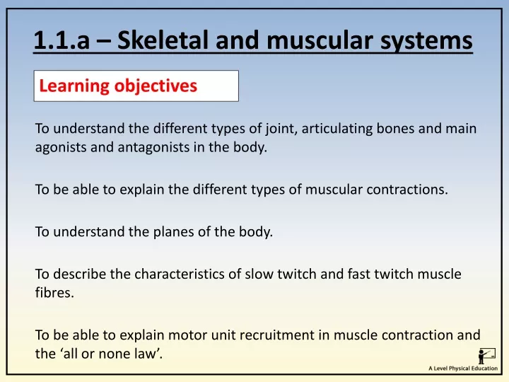
1.1.a – Skeletal and muscular systems Learning objectives To understand the different types of joint, articulating bones and main agonists and antagonists in the body. To be able to explain the different types of muscular contractions. To understand the planes of the body. To describe the characteristics of slow twitch and fast twitch muscle fibres. To be able to explain motor unit recruitment in muscle contraction and the ‘all or none law’.
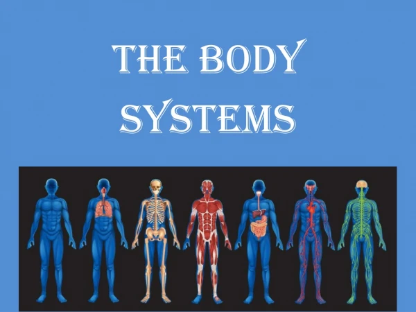
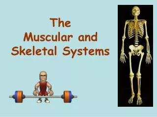
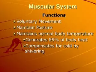
Skeletal System The skeleton is a framework for the body and provides protection, site for muscle attachment and is responsible for blood cell production. Watch me How many bone names do you know already?
Skeletal System Definition: “A joint is a place where two or more bones meet” The skeleton is a framework connected by joints. Joints are necessary for muscles to lever bones and create movement.
Skeletal System Head/Neck joint Cranium Vertebral column
Skeletal System Shoulder joint Scapula Humerus
Skeletal System Chest region Ribs Sternum
Skeletal System Elbow region Humerus Radius Ulna
Skeletal System Hip joint Femur Pelvis
Skeletal System Femur Patella Knee joint Tibia
Skeletal System Fibula Tibia Talus Ankle joint
Types of Joints Synovial joints are the most common type of joint in the body. These joints vary in structure for example, the shoulder is a ball-and-socket joint and the knee is a hinge joint. All synovial joints have the following structures. Synovial membrane Synovial fluid Joint capsule Ligament Cartilage
Types of Joints The following structures help prevent injury.
Types of Joints 1. Ball and socket joints allow movement in all directions and is the most mobile joints in the body. Examples: Shoulders and hips. Think. Pair. Share – Using examples, how are these joints used in sport? i.e. tennis serve
Classification of Joints 2. Hinge joints - only allow forwards and backwards movement like the hinge on a door. Examples found in the body: The knee and elbow. Why are these joints important for sport? These joint are extremely powerful and in conjunction with surrounding muscles can produce power and speed i.e. Knee drive during a 100m sprint
Classification of Joints 3. Pivot joints have a ring of bone that fits over a bone sticking out. Pivot joints allow rotation only. Examples found in the body: The joint between the atlas and axis in the neck which allows turning and nodding of head Why are these joints important for sport? This joint allows for small movements that assist a larger sporting action i.e. breathing during a swimming stroke
Classification of Joints 4. Condyloid joints have an oval-shaped bone end which fits into a similar shape. They allow small movement in all directions. Examples found in the body: Found between the carpals and metacarpals in the wrist joint. Why are these joints important for sport? These joint are extremely useful when a sport involves gripping a ball. i.e. handball throw
Classification of Joints 5. Gliding joints occur between the surfaces of two flat bones that are held together by ligaments. Examples found in the body: The bones in your wrists and ankles as well as the spine. Why are these joints important for sport? These joints are used to allow flexibility and movement in the hands, feet and back regions. i.e. a kicking or catching action or a boxing slip
Trapezius Muscle action External Obliques LatissimusDorsi Posterior Deltoid Pectolaris Major Teres Major Anterior Deltoid Triceps Brachii Gluteus Medius Biceps Brachii Wrist Flexors Gluteus Maximus Rectus Abdominals Wrist Extensors Adductor Longus Biceps Femoris Rectus Femoris Gastrocnemius Tibialis Anterior Soleus
Antagonistic muscle action Muscles are arranged in antagonistic pairs. As one muscle contracts (shortens) the other relaxes (lengthens). Think. Pair. Share - Can you think of another antagonists pair in the body?
Antagonistic muscle action Agonist – the contracting muscle responsible for causing movement. Antagonist – relaxing + lengthening muscle which allows the movement. (The muscle that works in opposition to the agonist) Antagonist (Biceps Brachii relax) Agonist (Tricep brachii contracts) Agonist (Biceps Brachii contract) Antagonist (Tricep brachii relax) Fixator – a muscle that stabilises one part of a body while the other moves.
Antagonistic muscle action Plantar Flexion - Gastrocnemius and Soleus (Agonist) and Tibialis Anterior (Antagonist) Flexion at the knee - Biceps Femoris (Agonist) and Rectus Femoris (Antagonist) Fixator - Gluteus Maximus
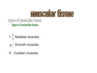
Types of muscular contractions • Isometric • Isotonic a. Concentric b. Eccentric
Types of muscular contractions Isometric contractions – These are muscle contractions that DO NOT create movement. Isometric contraction is when the muscle contracts without lengthening or shortening. The result is that no movement occurs. To hold the body in a particular position (e.g. scrum). Think. Pair. Share – Can you name any other sporting actions that are isometric?
Types of muscular contractions Isometric contractions happen when a movement is still/stationary or held.
Types of muscular contractions Isotonic contractions – A muscular contraction which changes the length of the muscle. This can occur in two ways; Concentric contractions – Concentric contraction is when the muscle shortens under tension. e.g. during the upward phase of an bicep curl, the biceps brachii performs a concentric contraction as it shortens to produce flexion of the elbow.
Types of muscular contractions - isotonic Eccentric contractions – Eccentric contraction is when the muscle lengthens under tension (and does not relax). When a muscle contracts eccentrically, it acts as a brake to help control the movement of the body part during negative work. e.g. when landing from a standing jump quadriceps muscles are contracting eccentrically.
Types of muscular contractions – Try this! Why not use some practical space and explore different muscular contractions and discuss whether they are Isometric or Isotonic, Concentric or Eccentric.
Movement analysis Flexion involves a decrease in the angle that occurs around a joint. i.e. radius and the humerus to decrease.
Movement analysis Extension involves an increasein the angle that occurs around a joint. i.e. straightening the elbow causes an increase in the angle between the humerus and the ulna/radius.
Movement analysis Planter Flexion is a term used solely for the ankle joint. It involves bending the foot downwards, away from the tibia. i.e. action of moving up onto toes or pointing toes.
Movement analysis Dorsi Flexion is bending the foot upwards towards the Tibia. i.e. - Action of pulling up toes towards the body.
Movement analysis Adduction - Movement towards midline of the body Abduction - Movement away from midline of the body
Movement analysis Horizontal flexion: Movement of the arm across the body in the horizontal (transverse) plane.
Movement analysis Horizontal extension: Movement of the arm away from the body in the horizontal (transverse) plane
Joints in action All sporting actions require different types of muscle contractions using a range of articulating bones, joints, movement patterns, agonist, antagonist and contraction types to perform the necessary movements. Articulating bones = Humerus/Ulna/Radius Type of Joint = Hinge Joint. Movement = Extension Agonist = Triceps BrachiiAntagonist = Biceps BrachiiContraction = Concentric Think. Pair. Share – Analyse the movement above at the elbow.
Joints in action Think. Pair. Share – Discuss and analyse the movements above.
The Shoulder joint The shoulder is a ball and socket joint where the head of the humerus fits into a cavity on the scapula. This type of joint allows the most movement. Its structure also makes it one of the least stable joints, so it is heavily reliant on ligaments and muscles to increase its stability.
The Shoulder joint The following muscles listed are the agonists responsible for the movement pattern. Anterior Deltoid (red) Posterior Deltoid (blue) Middle Deltoid (green) Latissimus Dorsi
The Shoulder joint Transverse Plane: Pectoralis Major Posterior Deltoid (blue)
The Shoulder joint Transverse Plane: Teres Major Subscapularis Teres Minor Infraspinatous
The elbow joint The elbow is a hinge joint, with the distal (far) end of the humerus articulating with the proximal (near) end of the radius and ulna. Movement can take place in one plane only, allowing only flexion and extension
The elbow joint Sagittal Plane: Biceps Brachii Triceps Brachii
The wrist joint The wrist is a condyloid joint, with the radius, ulna and carpals making up the joint. Wrist Flexors Wrist Extensors
The Hip joint The hip is a ball-and-socket joint where the head of the femur fits into the pelvic girdle. Action created by the Iliopsoas. Gluteus Maximus.
The Hip joint Adductor longus Adductor Brevis Adductor Magnus Gluteus Maximus Gluteus Minimus Gluteus Medius
The Knee joint The knee is classed as a hinge joint and allows flexion and extension only. Flexion: During the preparation for the action (backlift) the biceps femoris, semitendinous and semimembranosus concentrically contract. Extension: The downward kicking action involves the contraction of the rectus femoris, vastus lateralis, vastus intermedius and vastus medialis.
The Ankle joint The ankle is a hinge joint where the articulating bones are the tibia and fibula. The main muscles that control movement in this joint are the gastrocnemius, soleus and the tibialis anterior. These muscles allow plantarflexion and dorsiflexion movement.
Planes of movement To help explain movement, the body can be viewed as having a series of imaginary slices/glass panes running through it. These are referred to as planes of movement. For a movement to take place within a particular plane it must be parallel to that plane.
Planes 1.The sagittal plane is a vertical plane that divides the body into rightand leftsides. Think. Pair. Share – what joints in the body are capable of moving in the sagittal plane?
Sagittal Plane • The hinge joint is responsible for these movements. • Flexion and extension of the wrist, elbow, shoulder and knee. • Dorsi flexion and plantar flexion at the ankle.