Download
1 / 54
670 likes | 1.01k Views
T cell Development. Basic Principles of T Cell Development. Each cell randomly rearranges a specific TCR. The presence of the pMHC epitope, stage of development and “costimulation” determine the cell’s fate.

E N D
Basic Principles of T Cell Development • Each cell randomly rearranges a specific TCR. • The presence of the pMHC epitope, stage of development and “costimulation” determine the cell’s fate. • Functional Immunity & Tolerance result from the sum of a large population of many sequential pMHC epitope specific cellular responses.
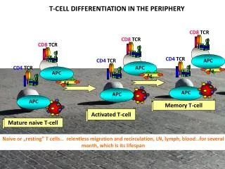
Following development into mature, antigen-responsive T cells, these T cells emerge from the thymus and migrate to secondary lymphoid tissues, where they interact with antigen, antigen-presenting cells, and other lymphocytes:
The importance of the thymus in T cell development is demonstrated by inherited immune deficiencies: people that do not have a thymus (DiGeorge’s syndrome, aka Thymic Aplasia) do not develop functional T cells. • DiGeorge’s syndrome results from a developmental defect – the failure of the third and fourth pharyngeal pouches to develop, which results not just in thymic defects, but also in absent parathyroids and in aortic arch defects. • Thymectomy early in life reduces the ability to produce T cells. • Thymectomy later in life does not markedly impair T cell number. • In fact, the thymus decreases in size with age. • However, the thymus can still produce new T cells up to middle-age, especially in situations where there is loss of T cells (HIV/AIDS).
While in the thymus, immature T cells, or thymocytes, undergo several changes that allow them to develop into mature T cells, ready for contact with antigen. • Thymocytes interact with thymic epithelial cells and various other cells while in the thymus.
The thymus is composed of several lobes, each of which has cortical and medullary regions:
The cortex contains immature thymocytes in close contact with thymic epithelial cells. • Medullary areas contain more mature thymocytes, epithelial cells, and dendritic cells and macrophages
During thymic differentiation, the great majority of thymocytes die by apoptosis, and are ingested by macrophages. • Only a small minority of these T cell progenitors make it out as mature T cells
Thymic development occurs in two phases: • production of T cell receptors for antigen, by rearrangement of the TCR genes 2) selection of T cells that can interact effectively with self-MHC
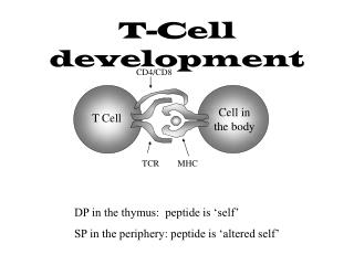
Changes in the expression of cell-surface molecules accompany the thymic differentiation of T cells: • entering thymocytes are TCR, CD3, CD4, and CD8-negative • as thymocytes mature, and undergo rearrangement of their TCR genes to generate a functional TCR, they begin to express CD3, CD4, and CD8 • mature T cells ready to go to the periphery are TCR/CD3+, and either CD4 or CD8 positive
First phase of thymic development: rearrangement of TCR genes to produce a functional TCR • Progenitor T cells enter the thymus (sub-capsular region of the outer cortex). • These cells do not have rearranged TCR genes and lack expression of characteristic T cell surface molecules. • Interaction with thymic stromal cells induces these progenitor T cells to proliferate. • These immature thymocytes do not yet express CD4 or CD8, molecules that are expressed by mature T cells: double-negative thymocytes.
There are two types of T cell receptors: gd andab • ab TCR T cells are the most abundant, by far: (or g & d chain)
Unlike B cells, in which the genes that encode the BCR rearrange in a set order, the TCR b, g, and d genes start to rearrange at about the same time. • If a productive g or d rearrangement occurs first, the T cell is committed to that lineage, and stops further rearrangement of the b TCR gene.
However, if b is rearranged first, then the T cell continues to proliferate, and undergoes further rearrangements. • This results either in rearranged a TCR gene, yielding an ab TCR lineage cell, or rearranging g and d genes, resulting in a gd TCR cell.
Rearrangements that lead to anab T cell begin the rearrangement of thebTCR gene. The first step is D-J joining, followed by VDJ rearrangement. Expression of b chain stops further b chain rearrangements.
b chain is then expressed on the surface of the thymocyte in association with a surrogatea chain (pTa). • Following this, there is rearrangement of thea TCR gene, resulting in a functional a chain, and in the expression of surface TCR, in association with other T cell-associated cell surface molecules.
During this process, a cell that makes an unproductive a chain rearrangement can try again until gets a good a chain, or it exhausts its possibilities:
Thymocytes that have a functional b rearrangement, and express ab or b + the surrogate a chain (pTa) are induced to express both CD4 and CD8 simultaneously – these are called double-positive cells. • Immature T cells that do not undergo a productive rearrangement die by apoptosis.
Second phase of thymic development: selection of T cells that can interact with self MHC and antigen • This applies only to ab TCR-bearing cells (>95% of T cells). • gd T cells are not restricted to interactions with MHC class I or class II molecules • This phase of T cell development consists of two steps: • positive selection (TCR that can interact withself-MHC) • negative selection (eliminate self-reactive cells that are stimulated by MHC + self)
Transgenic TCRs to Self AntigensClonal Deletion in Thymus • Double TCR transgenic mice (both TCR a and b) using a TCR from a CTL clone specific for male antigen (H-Y/H-2Db). • Female mice • most mature T cells expressed transgene TCR • normal proportion of CD4+ and CD8+ • 30% of CD8+ cells specific for male H-Y. • Male mice • large decrease (90%) in thymocytes (including CD4+8+) • down regulation of CD8+ among peripheral T cells • No H-Y reactive cells found; no autoimmunity P. Kisielow, H. Blüthmann, U. D. Staerz, M. Steinmetz, & H. von Boehmer. Nature 333:742-746 (1988)
Harold von Boehmer HY TCR transgenic mice C57BL/6 control male Expression of TCR Tg in females with H-2Db results in development of CD8+T (increased CD8/CD4) HY TCR transgenic Female Expression of TCR tg and autoantigen in males results in deletion of DP thymocytes and blocks development of Tg+ CD8 SP HY TCR transgenic male
11%; 2x10⁶ 5%; 1.5x10⁶ 0.6%; 1.2x10⁶ 101x106 91%; 9%; 2.7x10⁶ 26x106 75%; CD4 5.8%; 35x106 2.8%; 21x106 CD4 CD8 In Rag-/-HY TCR transgenics all thymocytes express the TCR tg. More dramatic skewing to CD8 in females anddeletion in males Female HY TCR tg Male HY TCR tg
Positive Selection • Positive selection refers to the selection of thymocytes that are able to bind to, and interact with, self-MHC molecules • In positive selection developing thymocytes continue to live if they bind MHC well enough to receive a signal through their TCR. • This signal is mediated by the interactions of these cells with MHC-expressing thymic cortical epithelial cells. • The ~95% of thymocytes that do not receive this signal undergo apoptosis.
Positive selection takes place in the cortex of the thymus lobules:
These CD4+ CD8+ TCR+ thymocytes interact with thymic epithelial cells that express both MHC class I and MHC class II molecules, complexed with self-peptides. • Thymocytes that bind MHC survive; those that don’t bind to self-MHC die. • TCR a chain rearrangements can continue during positive selection, allowing cells to explore alternative a chains for MHC binding. • Once a T cell is positively selected, TCR rearrangement stops.
Topi KO per MHC di classe II e transgenici per MHC classe II espressa esclusivamente a livello della regione corticale del timo sotto il controllo di un promotore specifico per i cheratinociti Selezione positiva di tutte le cellule T che riconoscono complessi MHC + peptide anche quelle fortemente autoreattive In assenza di presentazione dell’Ag a livello della midollare si ha selezione positiva incontrastata
Negative Selection • Negative selection refers to the elimination of those thymocytes that bind to self-MHC molecules + self with high affinity. • In negative selection developing thymocytes die if they bind MHC + self peptides too well (strongly enough so that they would be activated by this interaction, via signaling through their TCR).
Thymocytes undergo negative selection in the medullary region:
There, they interact with antigen-presenting cells (dendritic cells, macrophages) that express self-antigens + MHC class I or MHC class II molecules. • Thymocytes that bind to self + MHC too strongly are eliminated as possibly self-reactive cells, and undergo apoptosis. • If self-reactive T cells were allowed to exit the thymus, such cells would mediate autoimmune disease.
Effects of peptides acting as agonists, antagonists or neither on thymic selection