Download
1 / 47
480 likes | 610 Views
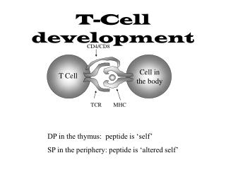
Two gene regulatory networks for T cell development. MBL GRN course Ellen Rothenberg Division of Biology, California Institute of Technology Oct. 18, 2011. The strategy of “adaptive immunity” and its developmental programming consequences .

E N D
Two gene regulatory networks for T cell development MBL GRN course Ellen Rothenberg Division of Biology, California Institute of Technology Oct. 18, 2011
The strategy of “adaptive immunity” and its developmental programming consequences • Every T or B cell has a “unique” antigen receptor recognition specificity via immune receptor (TCR or Ig) gene rearrangement and somatic mutation • Antigen receptor gene rearrangement process generates many dysfunctional products • Cells with bad receptor genes must be eliminated: >90% loss • Large excess of precursors must be generated continuously
The strategy of “adaptive immunity” and its developmental programming consequences: T cell development population dynamics Est. cell numbers: 1E07 3 – 50? 2E04 ~5E04 1E06 ~1E06 2E08 3E07
T cell development requires distinct program phases • Precursor expansion phase • Cell cycle arrest, TCR gene rearrangement, application of selection checkpoint (DN3 –|| b-selection) • Selected cell expansion phase • Cell cycle arrest, TCR gene rearrangement, application of selection checkpoint (DP –|| positive selection) • Distinct positive selection differentiation programs (helper, killer)
T cell development depends on extrinsic signals: crucial role of Notch/Delta signaling in vitro & in vivo Notch1-DL4 interaction is the major inductive signal that precursors receive from the thymus environment in vivo B cells T cells In vitro system: T. Schmitt & J. C. Zuniga-Pflucker, 2002 Rel. expression (Taghon et al., 2005, Genes Dev) Days
Notch-DL4 signalling drives and governs early stages of T-cell development but has hit & run role NOTCH requirement
Lockdown mechanisms in commitment • Positive regulatory factors for cell type • Expressed stably • Can act in positive feedback loops • Regulatory factors for alternative cell types • Must be silenced in development • May be repressed by lineage-specific factors • Continuous repression needed??? • Repression may be stabilized by chromatin closing
Genes in play during T-cell lineage commitment: a genome-wide look by RNA-seq(J. Zhang, A. Mortazavi, B. Williams, B.Wold, EVR) FLDN1 FLDN2a FLDN2b ThyDN3 ThyDP
T cell identity genes: details of gene-specific regulation differ …but many are strongly activated in DN2a-DN2b stage Data from RNA-seq, J. Zhang, A. Mortazavi, B. Williams, B. Wold, E. Rothenberg
“Lineage identity” factors: prototypes from other hematopoietic lineages Erythroid fate: GATA-1 B cell fate: EBF1 + Pax5 Myeloid fates: PU.1 + C/EBPa • Direct positive drivers of lineage-specific genes • Direct antagonists of alternative-lineage regulators • Direct antagonists of stem/progenitor cell genes • Lineage “specific” expression
Tcf7 (encodes TCF-1 protein) is induced by Notch/Delta signaling … Upregulation by Notch Upregulation by Tcf7 vs. Notch (Weber, … & Bhandoola, 2011, Nature)
Tcf7 is induced by Notch …and activatedTCF-1can turn on T-cell genes withoutNotch/Delta signaling Upregulation by Notch Upregulation by Tcf7 vs. Notch (Weber, … & Bhandoola, 2011, Nature)
Feed-forward network circuits triggered by Notch pathway signaling activate T-cell transcription factor genes and marker genes Notch signal Notch signal • Crucial role of Notch signal to activate: direct inputs to TCF-1 (Tcf7), Bcl11b, and some differentiation genes • Direct input from TCF-1 to Bcl11b and from GATA-3 to TCRb (& Cd3d,e) shown by ChIP-seq (Tcf7) • Dose dependence: • Normally activated Notch/Su(H) & sub-maximal Tcf7 collaborate to start specification • But maximal Tcf7 can turn on a subset of these targets without Notch input • This high-dose positive role of TCF is not dependent on Wnt/ b-catenin • (Weber et al., Nature 2011; P. Liu et al., Science, 2010; Wei et al., Immunity, 2011; J. Zhang et al., submitted; network models: H. Y. Kueh & EVR, WIREs Sys Bio, 2011)
Feed-forward network circuits triggered by Notch pathway signaling activate T-cell transcription factor genes and marker genes Notch signal Notch signal (Tcf7) • Crucial role of Notch signal to activate: direct inputs to TCF-1 (Tcf7), Bcl11b, and some differentiation genes • Direct input from TCF-1 to Bcl11b and from GATA-3 to TCRb (& Cd3d,e) shown by ChIP-seq • (Weber et al., Nature 2011; P. Liu et al., Science, 2010; Wei et al., Immunity, 2011; J. Zhang et al., submitted; network models: H. Y. Kueh & EVR, WIREs Sys Bio, 2011)
An unbiased look at transcription factor and DNA binding factor gene expression changes genome-wide Exp higher in DN1 Exp higher in DN2b PU.1 Log2 ratio DN1 exp: DN2b exp Gata3 Tcf7 Bcl11b
Major stages in early intrathymic T-cell development Major pathvia “DP” cells Bcl11b level
Conditional knockout of Bcl11b to determine its function in T-lineage commitment A. Bcl11b locus 3’UTR Exon 4 Loxp Loxp Conditional KO mouse model from Dr. Mark Leid, Oregon State University Infected by NGFR-Cre retrovirus (48 hr) B. NGFR+ c-kit+ CD27+ cells were sorted OP9-DL1 Flt3L, IL7 Fetal liver HSC: Lin-c-kit+ SCF, Flt3L, IL7 Cells were cocultured with OP9-DL1 for ~9 days. 1-2 weeks DN2A and DN2B cells were sorted Analysis, cell sorting OP9-DL1 SCF, Flt3L, IL7 (Long Li, Mark Leid, & E.V.R., Science 2010)
Loss of Bcl11b: deficient cells “stall” at early DN2a stage DN2 DN1 Sorted DN2a cells from Cre-treated: Bcl11b conditionalControl B6 miceknockout mice a c-Kit b DN4 DN3 further culture& differentiation CD25 Normal pathway for early T cell development DN3, DN4& later cells more DN2a cells& some NK cells(non-T) Bcl11b is needed to terminate “self-renewal” in DN2 stage (Long Li, Mark Leid, & E.V.R., Science 2010)
DN2a cells with deleted Bcl11b: “stuck” in DN2 but enhanced self-renewal capability Bcl11b-/-DN2A Bcl11b-/- “DN2B” B6 DN2A B6 DN2B c-kit CD25 Production of more DN2a cells in T-cell conditions Cell Numbers B6 DN2a + + Bcl11bKO DN2a + + Input 2 5 2 5
Early roles of Bcl11b (Long Li, Mark Leid, & E.V.R., Science 2010)
Molecular Basis of the Function of Bcl11b in T-cell Lineage Commitment • T-cell program • Stem Cell program • NK program (IL7Rα) CD3ε (initial) CD3γ CD25 Rag1 Lck GATA3 E2A HEB Tcf7 (TCF-1) Ikaros SCL (Tal1) Lyl1 Cpa3 Bcl11a Flt3 Gfi1b GM-CSFRβ Hhex Kit Sfpi1 (PU.1) (IL7Rα) Id2 IL2Rβ T-bet Eomes Zbtb16 (PLZF) Nfil3 (E4bp4) Zfp105 The initiation of T-lineage specification is Bcl11b-independent. The repression of the stem cell regulatory program requires Bcl11b. The repression of the NK program requires Bcl11b.
Bcl11b is not required to start the T-cell differentiation program
Bcl11b is the developmental gatekeeper for irreversibility of commitment
Factors expressed before commitment have “rap sheets” • Alternative lineage factors: PU.1 (myeloid, dendritic cell); Gfi1b, SCL/Tal1 (erythroid); Bcl11a, Lyl1 (B) • Stem cell self-renewal genes: SCL/Tal1, Lmo2, Erg, Hhex, Gfi1b, Meis1 • T-lineage proto-oncogenes: SCL/Tal1, Hhex, Lyl1, Lmo2
How simple a regulatory process is T-lineage commitment? • Use epigenetic markings to detect whencis-regulatory elements become active • H3K4me2 marks at promoter, enhancers, other functional sites • H3K(9,14)Ac marks closely correlate with pol II recruitment • When & how are regulatory genes that compete with commitment silenced • H3K27me3 marks silencing by PRC2 (Ezh2, Suz12) Analysis by ChIP-seq: same samples as used for RNA-seqtranscriptome analysis (J. Zhang, A. Mortazavi, B. Williams, B. Wold, & EVR)
H3K4me2 H3K(9,14)Ac H3K27me3 RNA All genes during T cell specification:Histone modifications at the transcriptional start site vs. RNA expression DN1, DN2a, DN2b, DN3, DP stages, left rightHeat map, log2 of integrated intensity
Lineage exclusion by repression of crucial regulators of other cell types • One or many different repression mechanisms required? • Temporally • Biochemically • When do these events occur – does this fit expectation for hierarchical order of changes in developmental potential? • Lose B cell potential earlier: EBF1, Pax5 • Lose DC, NK potential later: PU.1 (Sfpi1), Id2 • Stem cell self-renewal “potential”: SCL(Tal1), Lyl1, Erg, Gata2, Bcl11a, Hhex, Meis1 (Gfi1b)
Diverse repression mechanisms, not a single “master commitment factor”, silence regulators of different alternative fates: Pax5(B cell), Hhex (progenitor cell), Sfpi1/PU.1 (progenitors & myeloid cells) H3Ac H3K4me2 H3K27me3 RNA
Bcl11b knockout reveals a conditionally sustainable “Phase 1” state: what sustains it? E. V. Rothenberg, J. Zhang, L. Li 2010 Immunol Rev
An unbiased look at transcription factor and DNA binding factor gene expression changes genome-wide Exp higher in DN1 Exp higher in DN2b Three known roles: myeloid cells, B cells, multipotent progenitors PU.1 Log2 ratio DN1 exp: DN2b exp Gata3 Tcf7 Bcl11b
Impact on T cell development of removing PU.1 from DN1 stage: PU.1fl/fl FLPs OP9-DL1 co-culture OP9-DL1 co-culture 3 days 4 hrs Infection with Cre-carrying retroviruses e14.5 B6 and PU.1fl/fl FL cells 2 days Sort Cre+ DN1, DN2a and DN2b cells Conditional knockout mouse model from Dr. Stephen Nutt, WEHI DN progression
Accelerated T-cell differentiation in the absence of PU.1, but reduced cell recovery: tradeoff between phase 1 proliferation and phase 2 differentiation Ameya Champhekar
Two “phase 1” transcription factor genes with PU.1 binding sites are functional positive regulatory targets of PU.1 Upregulated in cells forced to express wildtype PU.1 (PU.1-WT) Downregulated in cells forced to express an artificial obligate repressor form of PU.1 (PU.1 Eng) (RNA expression rel. to b-actin, avg + SD, by qPCR) (Ameya Champhekar)
PU.1 has direct inputs into multiple phase 1 specific genes PU.1 binding targets Lmo1, Lmo2, (Tal1), Lyl1, Hhex, and Bcl11a are all proto-oncogenes PU.1 Sfpi1 Lyl1 Meis1 Flt3 Bcl11a Lmo2 Hhex PU.1 binding at promoters and specific distal elements defined by ChIP-seq; regulatory impact of inputs defined by gain/loss of PU.1 function effects (qPCR)
Phase 1 and phase 2 positive networks: what is their interaction? PU.1 Sfpi1 Lyl1 Meis1 Flt3 Bcl11a Lmo2 Hhex ? ? ? ? Notch signal Notch signal
PU.1 overexpression represses T-cell specific genes: a potential timing role for completion of commitment But does it do this directly or via activation of another phase 1 repressor? Strategy: use modified PU.1 gene with obligate repressor activity
PU.1-Engrailed fusion construct: test for PU.1 functional role by forced expression of obligate repressor PU.1 Transactivation DNA binding HDAC Repression of PU.1 target genes Gro/TLE
Control PU.1-WT PU.1-Eng Log10expression DN2b DN2b DN2b DN2a DN2a DN2a DN1 DN1 DN1 Stem cell and Alt. lineage genes PU.1 overexpression blocks or delays activation of specific T-cell genes in early T lineage cells T-cell genes
Control PU.1-WT PU.1-Eng DN2b DN2b DN2b DN2a DN2a DN2a DN1 DN1 DN1 Stem cell and Alt. lineage genes But…PU.1 repression of T-cell genes is probably indirect Form with obligate repressor biochemical action represses true PU.1 activation targets but activates T-cell genes T-cell genes
PU.1 overexpression represses T-cell genes through activating another negative regulator Positive regulators Positive regulators Upregulation by PU.1-Eng implies that endogenous PU.1 in vivo counterbalances action of positive drivers (e.g. Notch, GATA3, TCF-1, E proteins) that are already expressed before commitment
A regulated phase 1-phase 2 transition is required: “phase 1” factors as well as “phase 2” are part of T-cell program Thymus Stem cell Multipotent “Paucipotent” Committed Notchsignals ETPDN1 DN2a DN2b DN3a Grow! Restrain growth; make TCR ROLE IN STAGE: KEY FACTORS: “Stem/progenitor” PU.1, Lyl1, SCL, Hhex,Gfi1b, Erg, Bcl11a… T-cell factors…?
Transcription factor expression: multiple patterns from multipotent stage to T-cell lineage commitment Rising Falling DN3 peak Legacy drop Genes from gene discovery project (David-Fung et al., Devel. Biol., 2009); levels measured by qPCR (log10 scale); patterns identified by supervised clustering (self-organizing matrices)
T cell development depends on extrinsic signals: crucial role of Notch/Delta signaling in vitro & in vivo B cells Tcf7=TCF-1 T cells Rel. expression (Taghon et al., 2005, Genes Dev) Days
T-cell development depends on transcription factors that are shared with other programs Implication: Not just presence of factor, but its network connections and stage of action determines T-lineage role