Download
1 / 53
530 likes | 535 Views
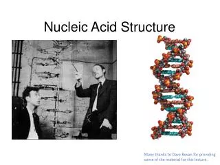
Nucleic Acid Structure and Stability. Chapter 8 (Page 283-302). Secondary Level of Nucleic Acid Structure (DNA). 1 . Watson-Crick Model of DNA. 1I. Replication based on Watson-Crick Model of DNA. Strand separation occurs first.

E N D
Nucleic Acid Structure and Stability Chapter 8 (Page 283-302)
Secondary Level of Nucleic Acid Structure (DNA)
1I. Replication based on Watson-Crick Model of DNA Strand separation occurs first. Each strand serves as a template for the synthesis of a new strand specified by the base-pairing rules. Synthesis is catalyzed by enzymes known as DNA polymerases. Newly made DNA molecule has one daughter strand and one parent strand.
2. Structural Variants of DNA There are two main classes of DNA structural variations. Different three-dimensional forms due to nucleotide conformations. Unusual DNA sequence structures.
2I. DNA Structural Variations arising from Nucleotide Conformation The conformation of a nucleotide in DNA is affected by rotation about seven different bonds. Restricted rotation around bond 4 results in ring-puckering (endo and exo conformations). Rotation around bond 7 results in different 3-D structures.
2Ib. Conformation of the Nucleobases. For purine bases, only two conformations with respect to the attached ribose units are sterically permitted, anti and syn. Pyrimidines generally occur in the anti conformation due to steric hindrance between C-2 of the pyrimidine and the sugar. 2
2Ic. A, B, and Z forms of DNA. • The Watson-Crick structure is referred to as the B-form DNA. This form is the most stable structure for a random-sequence DNA molecule under physiological conditions.
2Id. A form of DNA. • The right-handed A form is favored in nonaqueous solutions. • Most DNA crystallize in this form due to dehydration from crystallization methods. • The helix is wider than the B form and has 11 base pairs per helical turn (rather than 10.5). • The plane of the base pairs is tilted about 20° with respect to the helix axis. • Deeper major groove • Shallower minor groove
2Ie. Z form of DNA. The backbone of the left-handed helix takes on a zigzag appearance and the structure is more slender and elongated. There are 12 base pairs per helical turn. Prominent form when pyrimidines alternate with purine, especially alternating C and G. The purine adopts a syn conformation while the pyridimine is in the anti conformation. The major groove is barely apparent but minor groove is deepen yet narrow. Does exist in vivo though not abundantly.
2II. Unusual DNA Sequence Structures • Palindrome (word/phrase spelled identically read either forward or backward) • Sequence of double-stranded nucleic acid with inverted repeats of base sequence having twofold symmetry. • The sequence is superimposed by rotating 180° about the horizontal axis then 180° about the vertical axis.
2II. Unusual DNA Sequence Structures • Such sequences are self-complementary within each strand and can form a hairpin
2II. Unusual DNA Sequence Structures • Or form a cruciform with two strands
2II. Unusual DNA Sequence Structures • B. Mirror repeat • Has a symmetric sequence within each strand. • Involves superimposition of one repeat on the other repeat by a single 180° rotation about the vertical axis. • Does not have complementary sequences within the same strand and can not form hairpins or cruciforms.
2II. Unusual DNA Sequence Structures • Present in H-DNA, a triplex DNA structure.
2II. Unusual DNA Sequence Structures • C. Triplex DNA • This structure appears during important DNA metabolic events (replication, recombination, transcription). • It contains a polypyrimidine or polypurine tract. • - Can consist of two pyrimidine strands with one purine strand or two purine strands with one pyrimidine strand. • Involves parallel and antiparallel strand base pairing.
2II. Unusual DNA Sequence Structures • Hoogsteen pairing of the nucleobases occurs in these structures.
2II. Unusual DNA Sequence Structures • D. G tetraplex • Four-stranded helix • This structure occurs readily for DNA sequences with a very high proportion of guanosine residues. • The orientation of the strands can be antiparallel or all parallel.
Secondary Level of Nucleic Acid Structure (RNA)
1. Single-stranded RNA Transcription Translation DNA RNA Protein Replication • The product of DNA transcription is always single-stranded RNA. RNA does not have a simple, regular secondary structure. Generally, • Right-handed helix dominated by base-stacking interactions. • Base-stacking is strongest between two purines. • A pyrimidine in between two purines is often displaced to enable the two purines to interact.
2. RNA can form a Double-Helix • RNA can base-pair with complementary regions of either RNA or DNA. • G-C • A-U (or the occasional T) • G-U (fairly common but a folding phenomenon) • The RNA double-helixtakes on the A-form right-handed double helix. • B or Z forms have not been observed in vivo.
2. RNA can form a Double-Helix • Breaks in the A-form helix caused by mismatched or unmatches bases in one or both strands are common and result in bulges, internal loops, and hairpen loops. Hairpen Loop Internal Loop Bulge
3. RNA can form Complex Structures • There are extensive base-paired helical structures in many RNAs. • The hairpins that result from these structures are the most common type of secondary structure in RNA.
Tertiary Level of Nucleic Acid Structure (DNA)
1. Circular vs Linear DNA • DNA strands exist in double-helical structures in two forms: • Circular (Circular DNA) as is common in viruses, bacteria, and archaea. This form is also present in eukaryotic cells. • Linear (Linear DNA), which is the dominant form in humans. Circular DNA Linear DNA
2. DNA Supercoiling • Cellular DNA is extremely compacted and requires a high degree of structural organization. • The folding mechanism must not only pack the DNA but also permit access to the information in the DNA. • The compact structure is achieved by supercoiling. Relaxed State
3. Compaction of DNA in a Eukaryotic Chromosome • The chromosome is a single large DNA molecule and its associated proteins, which stores and transmits genetic information.
4. Agarose Gels to Visualize DNA • Agarose gel electrophoresis is a method to determine the size of DNA but can also provide insight into DNA forms. • DNA migrates through a highly cross-linked agarosematrix in response to an electric field. • The phosphates on the DNA are negatively charged and the molecule migrates to the positively charged electrode. • Three factors affect migration rate • Size of the DNA (measured in # of base pairs (BP)) • Conformation of the DNA • Ionic strength of the running buffer
4. Agarose Gels to Visualize DNA MW Ladder DNA is typically visualized via ethidium bromide (EtBr) staining. - EtBr is a DNA intercalator.
4. Agarose Gels can Distinguish DNA Forms • DNA conformation will affect migration rate through agarose gels. • Relaxed forms of DNA travel more slowly through the gel. With the circular form traveling slower than linear form. • Supercoiled form travels the fastest. • A restriction enzyme can be used to cut DNA at a specific site to linearize it.
5. DNA Isolation, Purification, and Identification • Unlike protein isolation and purification, the process is more straightforward for DNA samples • Standard centrifugation protocols can be used to isolate high quality DNA samples from organisms (lysates). • DNA isolation can be confirmed by agarosegelsand the DNA can be purified from the gels using standard kits. • DNA sequence can be identified by traditional protocols such as the Sanger method. • DNA cloning can be used to amplify DNA content.
1. Inherent Stability of DNA • The role of DNA as a repository of genetic information depends on its inherent stability. This stability can be evaluated in terms of: • Denaturation • Nonenzymatic Transformations • Very slow reactions can have a significant affect on DNA structure/function
2. DNA Denaturation • Denaturation can be induced by high temperature or change in pH.The process is not uniform and involves: • Covalent bonds remaining intact • Therefore the genetic code remains intact • Hydrogen bonds breaking • This results in the two strands separating • Disruption of base stacking • The close interaction between stacked bases of stable DNA interferes with their UV absorbing abilities leading to a hypochromic effect- a decrease in their absorption • Denaturation results in an increase in the UV absorbance (hyperchromic effect)
2. DNA Denaturation • D. Renaturation is the reverse of denaturation • Arapid one-step process called annealingas long as part of the double-strand remains intact. • If the double-strand is not intact then the process is two-step, with the first being a relatively slow step in which complementary strand pairing occurs via random collisions.
2I. Thermal DNA Denaturation (Melting) • DNA exists as a double helix at physiological temperatures. • The two DNA strands dissociate at elevated temperatures. • The strands re-anneal when temperature is lowered. • The reversible thermal denaturation and annealing form basis for the polymerase chain reaction. • DNA denaturation is commonly monitored by UV spectroscopy at 260 nm. • RNA duplexes and RNA-DNA hybrids can also be denatured.
2II. Factors Affecting DNA Denaturation • A. The midpoint of melting (Tm)depends on DNA length • Longer DNA has higher Tm • B. Tmdepends on pH and ionic strength • High salt increases Tm • C. Tmdepends on base composition • High CG increases Tm • AT rich regions melt first
2III. DNA Hybridization via Renaturation • The ability of two complementary DNA strands to pair with one another as DNA hybrids • Reveals evolutionary relationships • The closer the evolutionary relationship between two species, the more extensively their DNAs will hybridize • Enables modern molecular genetics techniques • Polymerase chain reaction • Site-directed mutagenesis
3. Nonenzymatic Transformations • Spontaneous Mutagenesis: • A. Deamination • Very slow reactions • Large number of residues • The net effect is significant: 100 C U events /day in a mammalian cell • B. Depurination • N-glycosidic bond is hydrolyzed • Significant for purines: 10,000 purines lost/day in a mammalian cell • Fortunately, cells have mechanisms to correct most of these modifications.
3. Nonenzymatic Transformations Radiation-Induced Mutagenesis: A. UV light induces dimerization of pyrimidines, which cause kinks in DNA structure. This effect isthe main mechanism for skin cancers. B. Ionizing radiation (X-rays and -rays) causes ring opening and strand breaking . Cells can repair some of these modifications, but others cause permanent and detrimental mutations. Accumulation of these type of mutations is linked to aging and carcinogenesis.