Download
1 / 46
460 likes | 464 Views
Explore the way signals pass across membranes, ion channels and the chemistry of nucleic acids. Topics include G proteins, adenylyl cyclase, inositol-phospholipid signaling pathway, receptor tyrosine kinases, ion channels, and nucleotide bases.

E N D
Membrane signaling; nucleic acid structure Andy HowardIntroductory BiochemistryWednesday 15 October 2014 Membrane Signaling; Nucleic Acids
Membranes: transport and signaling • We’ll talk about the way that signals pass across membranes and deepen our understanding of ion channels; • Then we’ll introduce the chemistry and structure of nucleic acids Membrane Signaling; Nucleic Acids
Signal transduction G proteins Adenylyl cyclase Inositol-phospholipid signaling pathway Sphingolipid messages Receptor tyrosine kinases Ion channels General principles Modes of gating Nucleotide bases Pyrimidines Purines Tautomers Nucleosides & -tides Deoxynucleosides What we’ll discuss Membrane Signaling; Nucleic Acids
G proteins (G&G § 32.4) • Transducers of external signals into the inside of the cell • These are GTPases (GTP GDP + Pi) • GTP-bound protein transduces signalsGDP-bound protein doesn’t • Heterotrimeric proteins; association of b and g subunits with a subunit is disrupted by complexation with hormone-receptor complex, allowing departure of GDP & binding of GTP Membrane Signaling; Nucleic Acids
GTP Inactive GDP G protein cycle a Active b a g GTP b • Ternary complex disrupted by binding of receptor complex • Ga-GTP interacts with effector enzyme • GTP slowly hydrolyzed away • Then Ga-GDP reassociates with b,g H2O g Pi a GDP Inactive Membrane Signaling; Nucleic Acids
Adenylyl cyclase • cAMP and cGMP:second messengers • Adenylyl cyclase convertsATP to cAMP • Integral membrane enzyme;active site faces cytosol • cAMP diffuses from membrane surface through cytosol, activates protein kinase A • Protein Kinase A (PKA) phosphorylates ser,thr in target enzymes; action is reversed by specific phosphatases Cyclic AMP Membrane Signaling; Nucleic Acids
Modulators of cAMP • Caffeine, theophylline inhibit cAMP phosphodiesterase, prolonging cAMP’s stimulatory effects on protein kinase A • Hormones that bind to stimulatory receptors activate adenylyl cyclase, raising cAMP levels • Hormones that bind to inhibitory receptors inhibit adenylyl cyclase activity via receptor interaction with the transducer Gi. Membrane Signaling; Nucleic Acids
Inositol-Phospholipid Signaling Pathway PIP2 • 2 Second messengers derived from phosphatidylinositol 4,5-bisphosphate (PIP2) • Ligand binds to specific receptor; signal transduced through G protein called Gq • Active form activates phosphoinositide-specific phospholipase C bound to cytoplasmic face of plasma membrane Membrane Signaling; Nucleic Acids
PIP2 chemistry • Phospholipase C hydrolyzes PIP2 to inositol 1,4,5-trisphosphate (IP3) and diacylglycerol • Both of these products are second messengers that transmit the signal into the cell Membrane Signaling; Nucleic Acids
IP3 and calcium • IP3 diffuses through cytosol and binds to a calcium channel in the membrane of the endoplasmic reticulum • The calcium channel opens, releasing Ca2+ from lumen of ER into cytosol • Ca2+ is a short-lived 2nd messenger too: it activates Ca2+-dependent protein kinases that catalyze phosphorylation of certain proteins Membrane Signaling; Nucleic Acids
Calcium homeostasis & IP3 Courtesy Oulu Univ., Finland Membrane Signaling; Nucleic Acids
Diacylglycerol and protein kinase C • Diacylglycerol stays @ plasma membrane • Protein kinase C (which exists in equilibrium between soluble & peripheral-membrane form) moves to inner face of membrane; it binds transiently and is activated by diacylglycerol and Ca2+ • Protein kinase C catalyzes phosphorylation of several proteins Membrane Signaling; Nucleic Acids
Control of inositol-phospholipid pathway Figure courtesyMotifolio.com • After GTP hydrolysis, Gq is inactive so it no longer stimulates Plase C • Activities of 2nd messengers are transient • IP3 rapidly hydrolyzed to other things • Diacylglycerol is phosphorylated to form phosphatidate Membrane Signaling; Nucleic Acids
The big picture Courtesy bmj.com Membrane Signaling; Nucleic Acids
Sphingolipids give rise to 2nd messengers • Some signals activate hydrolases that convert sphingomyelin to: • sphingosine • sphingosine-1-P, and • ceramide • Each of these modulates a second messenger Membrane Signaling; Nucleic Acids
Interconversions Courtesy AOCS Lipid Library Membrane Signaling; Nucleic Acids
Fates of sphingolipid products • Sphingosine inhibitsProtein Kinase C • Ceramides activate a protein kinase and a protein phosphatase • Sphingosine-1-P can activate Phospholipase D, which catalyzes hydrolysis of phosphatidylcholine;products are 2nd messengers Phospholipase DStreptomyceswith phosphatidyl choline boundPDB 2ZE454 kDa monomer2.5Å Membrane Signaling; Nucleic Acids
ligands exterior Receptor tyrosine kinases Tyr kinase monomers interior • Most growth factors function via a pathway that involves these enzymes • In absence of ligand, 2 nearby tyr kinase molecules are separated • Upon substrate binding they come together, form a dimer Membrane Signaling; Nucleic Acids
Autophosphorylation of the dimer P P • Enzyme catalyzes phosphorylation of specific tyr residues in the kinase itself; so this is autophosphorylation • Once it’s phosphorylated, it’s activated and can phosphorylate various cytosolic proteins, starting a cascade of events Membrane Signaling; Nucleic Acids
Insulin receptor • Insulin binds to an a2b2 tetramer;binding brings b subunits together • Each tyr kinase (b) subunit phosphorylates the other one • The activated tetramer can phosphorylate cytosolic proteins involved in metabolite regulation Sketch courtesy ofDavidson College, NC Membrane Signaling; Nucleic Acids
Ion channels: general principles (G&G §9.7) • Channels are selective for particular ions • Gated—i.e. they can open or close in response to specific signals • Voltage-gated: open or close in response to change in membrane potential • Ligand-gated: open or close in response to binding of an ion, a small organic molecule, or a protein Membrane Signaling; Nucleic Acids
General principles II • The opening through which the ion will flow is typically lined with charged amino acids (- for + ion, + for - ion) • Ions may or may not be hydrated as they flow Membrane Signaling; Nucleic Acids
Potassium channels • Important because a lot of cells depend on K+ for various cellular processes • Conduct K+ at close to diffusion rates • When open, they allow K+ but not Na+ to pass through based on ionic radius (1.33Å vs. 0.95Å) Rod MacKinnon Membrane Signaling; Nucleic Acids
KcsA from Streptococcus lividans • Homotetramer • Water-filled pore that goes >halfway through the bilayer,ending at the filter • In the crystal structure:Hydrated K+ ion suspendedin the center • Each subunit contributes 3 helices • Selectivity filter built from one pentapeptide (TVGYG) loop per subunit • Cf. G&G fig. 9.40 KcsA + Fab56 kDa tetramer;monomer+Fab shown PDB 1R3J, 1.9Å Membrane Signaling; Nucleic Acids
Gating in KcsA • Opening of channel is gated by intracellular pH:Closed at neutral pH, opened at acidic pH • Helix bending & rearrangement enable the opening up of the pore (G&G fig. 9.42) • 4 sites: typically 2 contain K+, 2 contain water Figure from Thompson et al. (2008) PNAS 105: 6900-6905. Membrane Signaling; Nucleic Acids
Gating in other K+ channels • Mammalian K+ channels are often voltage gated • Helix S4 has 5 Arg facing outside surface of membrane; • Closing the channel involves moving S4 to push down on the linker, inducing a change in S5 & S6, closing the channel • Closed @ resting potential of -60 mV; open ~ +30 mV • Note: PDB entry in fig. 9.43 is wrong. 1J4C doesn’t exist; 1K4C is the bacterial K+ channel Membrane Signaling; Nucleic Acids
Other selectivity filters • Replace TVGYG with TVGDG and you get an NaK channel • Similar to transient receptor potential (TRP) channels • Cf. G&G fig. 9.45 Bacillus cereusNaK channel24.9kDa dimerPDB 2AHY, 2.4Å Membrane Signaling; Nucleic Acids
CorA: a pentameric Co2+/Mg2+ channel • Bacterial, archaeal Mg2+transport • Big cytosolic N-terminal domain,C-terminal transmembrane domain • More than 100Å long helix! • Many fivefold-symmetric relationships define the pore • Long helix and the negatively-charged helices might act as a lever to open the pore • This channel is substrate (and potential)-gated. Thermatoga CorAPDB 4I0U, 2.7Å209kDa pentamer Membrane Signaling; Nucleic Acids
Single-subunit channels(G&G fig. 9.47) • ClC channels are homodimers • each monomer can transport Cl- • 3 Cl- sites; high [Cl-] displaces a glu near the extracellular medium, allows escape • AmtB: trimeric NH4+ transporter;converts ion to neutral ammonia as it passes • GlpF: Tetrameric glycerol channel: gradually dehydrates its substrates as it moves them • Apq1: Aquaporin water channels like GlpF Spinachaquaporin130 kDa tetramer, monomer shownPDB 3CN5, 2.1Å Membrane Signaling; Nucleic Acids
Chemistry Nobel Prize 2009 • Structural studies of the ribosome • Venki Ramakrishnan, LMB Cambridge • Thomas Steitz, HHMI Yale University • Ada Yonath, Weizmann Institute Membrane Signaling; Nucleic Acids
6 1 5 Pyrimidines 4 2 3 • Single-ring nucleic acid bases • 6-atom ring; always two nitrogens in the ring, meta to one another • Based on pyrimidine, although pyrimidine itself is not a biologically important molecule • Variations depend on oxygens and nitrogens attached to ring carbons • Tautomerization possible • Note line of symmetry in pyrimidine structure Membrane Signaling; Nucleic Acids
Uracil and thymine • Uracil is a simple dioxo derivative of pyrimidine: 2,4-dioxopyrimidine • Thymine is 5-methyluracil • Uracil is found in RNA; Thymine is found in DNA • We can draw other tautomers where we move the protons to the oxygens Membrane Signaling; Nucleic Acids
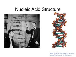
Tautomers • Lactam and Lactim forms • Getting these right was essential to Watson & Crick’s development of the DNA double helical model Membrane Signaling; Nucleic Acids
Cytosine • This is 2-oxo,4-aminopyrimidine • It’s the other pyrimidine base found in DNA & RNA • Spontaneous deamination (CU) • Again, other tautomers can be drawn Membrane Signaling; Nucleic Acids
Cytosine:amino and imino forms • Again, this tautomerization needs to be kept in mind Membrane Signaling; Nucleic Acids
7 6 5 1 8 4 Purines 2 9 3 • Derivatives of purine; again, the root molecule isn’t biologically important • Six-membered ring looks a lot like pyrimidine • Numbering works somewhat differently: note that the glycosidic bonds will be to N9, whereas it’s to N1 in pyrimidines Membrane Signaling; Nucleic Acids
Adenine • This is 6-aminopurine • Found in RNA and DNA • We’ve seen how important adenosine and its derivatives are in metabolism • Tautomerization happens here too Membrane Signaling; Nucleic Acids
Guanine • This is 2-amino-6-oxopurine • Found in RNA, DNA • Lactam, lactim forms Membrane Signaling; Nucleic Acids
Other natural purines • Hypoxanthine and xanthine are biosynthetic precursors of A & G • Urate is important in nitrogen excretion pathways Membrane Signaling; Nucleic Acids
Tautomerization and H-bonds • Lactam forms predominate at neutral pH • This influences which bases are H-bond donors or acceptors • Amino groups in C, A, G make H-bonds • So do ring nitrogens at 3 in pyrimidines and 1 in purines • … and oxygens at 4 in U,T, 2 in C, 6 in G Membrane Signaling; Nucleic Acids
Nucleosides • As mentioned in ch. 7, these are glycosides of the nucleic acid bases • Sugar is always ribose or deoxyribose • Connected nitrogen is: • N1 for pyrimidines (on 6-membered ring) • N9 for purines (on 5-membered ring) Membrane Signaling; Nucleic Acids
Pyrimidine nucleosides • Drawn here in amino and lactam forms Membrane Signaling; Nucleic Acids
Pyrimidine deoxynucleosides Membrane Signaling; Nucleic Acids
A tricky nomenclature issue • Remember that thymidine and its phosphorylated derivatives ordinarily occur associated with deoxyribose, not ribose • Therefore many people leave off the deoxy- prefix in names of thymidine and its derivatives: it’s usually assumed. Membrane Signaling; Nucleic Acids
Purine nucleosides • Drawn in amino and lactam forms Membrane Signaling; Nucleic Acids
Purine deoxynucleosides Membrane Signaling; Nucleic Acids