Download
1 / 29
290 likes | 418 Views
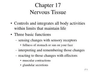
Chapter 17 The Nervous System. The Brain 4 regions: cerebrum, diencephalon, brainstem, cerebellum Contains interconnecting neurons (cell bodies and axons) Gray matter: aggregations of neuronal cell bodies White matter: neuronal axons coated with myelin.

E N D
The Brain 4 regions: cerebrum, diencephalon, brainstem, cerebellum Contains interconnecting neurons (cell bodies and axons) Gray matter: aggregations of neuronal cell bodies White matter: neuronal axons coated with myelin Central and Peripheral Nervous System — Key Definitions Central nervous system: the brain and spinal cord
The spinal cord Extends from brainstem (medulla) to L1-L2 vertebrae Contains motor and sensory pathways that exit and enter the cord via anterior and posterior nerve roots and spinal and peripheral nerves 5 segments: cervical (C1-8), thoracic (T1-12), lumbar (L1-5), sacral (S1-5), coccygeal Central Nervous System – Brain and Spinal Cord Note: Cauda equina at L1-2, where nerve roots fan out like a horse’s tail
Peripheral Nervous System – Cranial Nerves • Peripheral nervous system • 12 pairs of cranial nerves plus spinal and peripheral nerves • Cranial nerves govern motor, sensory, and specialized functions like smell, vision, and hearing
Peripheral Nervous System – Peripheral Nerves • Peripheral nerves: 31 pairs of nerves that attach to the spinal cord: 8 cervical, 2 thoracic, 5 lumbar, 5 sacral, 1 coccygeal • Each nerve has an anterior (ventral) root containing motor fibers and a posterior (dorsal) root containing sensory fibers; the anterior and posterior roots merge to form a short (<5 mm) spinal nerve • Spinal nerve fibers commingle with similar fibers from other levels to form peripheral nerves
Peripheral Nervous System — Motor and Sensory Pathways and Dermatomes • Motor and sensory pathways: descending motor and ascending sensory pathways • Dermatome: band of skin innervated by the sensory root of a single spinal nerve
Common or Concerning Symptoms of the Nervous System • Headache • Dizziness or vertigo • Generalized, proximal, or distal weakness • Numbness • Abnormal or loss of sensations • Loss of consciousness, syncope, or near-syncope • Seizures • Tremors or involuntary movements
Heath Promotion and Counseling • Preventing stoke orTIA • Reducing risk of peripheral neuropathy • Detecting the “three Ds” – delirium, dementia, and depression
The Nervous System: Key Principles • As you examine the patient, remember three important questions: • Is mental status intact? • Are right- and left-sided findings the same, or symmetric? • If findings are asymmetric or otherwise abnormal, do the causative lesions lie in the central nervous system or the peripheral nervous system? • Organize your thinking into 5 categories: mental status, speech, and language; cranial nerves; motor system; sensory system; and reflexes
Examination – Motor System • Position, movement, muscle bulk, and tone • Observe body position and involuntary movements such as tremors, tics, fasciculations • Inspect muscle bulk; note any atrophy • Assess muscle tone — flex and extend the arm and the lower leg for residual tension → slight resistance to passive stretch
Examination – Muscle Strength • Ask the patient to move actively against your opposing resistance; assign Grade 5 if the patient overcomes your opposing movement • If the patient can only move against gravity, assign Grade 3
Examination – Muscle Strength (cont.) • Test the following muscle groups and movements: • Biceps and triceps, wrist – flexion and extension • Handgrip, finger – abduction and adduction, thumb opposition • Trunk – flexion, extension, lateral bending • Thorax – expansion, diaphragmatic excursion during respiration • Hip – flexion, extension, abduction, and adduction • Knee and ankle – flexion, extension
Examination – Coordination • Test coordination, including: • Rapid alternating movements – patient turns hand rapidly over and back on thigh; taps tip of index finger rapidly on distal thumb; taps ball of foot rapidly on your hand • Point-to-point movements – patient touches nose then your index finger as you move it to different positions; patient moves heel from opposite knee down the shin to the big toe • Gait – assess gait as patient: • Walks across room • Walks heel-to-toe • Walks on toes then heels • Hops in place
Question Coordination of muscle movement requires that four areas of the nervous system function in an integrated way. Coordinating eye, head, and body movements applies to which area of the nervous system? • Motor system • Cerebellar system • Vestibular system • Sensory system
Answer • Vestibular system: balance and coordinating eye, head, and body movements • Motor system: muscle strength • Cerebellar system: rhythmic movement and steady posture • Sensory system: position sense
Examination – Coordination (cont.) • Test coordination, including: • Stance, namely: • The Romberg test • Patient stands with feet together and eyes open, then with eyes closed for 30–60 seconds without support • Loss of balance when eyes closed is a positive test • Pronator drift • Patient stands for 20–30 seconds with both arms straight forward, palms up, and eyes closed; tap arms briskly downward • Pronation and downward drift of the arm is a positive test
Examination – Sensory System: General Principles • Compare symmetric areas on both sides of the body • When testing pain, temperature, and touch, compare distal with proximal areas of the extremities • Map out the boundaries of any area of sensory loss or hypersensitivity
Examination – Sensory System • Test pain: use a disposable object such as a broken cotton swab or pin and discard after each use. • Ask if prick is sharp or dull, or ask the patient to compare 2 sensations: “Does this feel the same on both sides?” • Test light touch, using cotton wisp. • Test vibration: tap a 128-Hz tuning fork on your hand, then place it on the DIP joint of the patient’s finger. Ask the patient, “Do you feel a buzz? Tell me when it stops.” Likewise test over the joint of the big toe. • Test proprioception: hold the big toe by its sides between your thumb and index finger, pull it away from the other toes, and move it up then down. Ask the patient to identify the direction of movement.
Examination – Sensory System (cont.) • Assess discriminative sensation to test the ability of the sensory cortex to analyze and interpret sensations • Stereognosis: place a key or familiar object in the patient’s hand and ask the patient to identify it • Number identification (graphesthesia): outline a large number in the patient’s palm and ask the patient to identify the number • Two-point discrimination: using two ends of an opened paper clip, or two pins, touch the finger pad in two places simultaneously; ask the patient to identify 1 touch or 2 • Point localization: lightly touch a point on the patient’s skin and ask the patient to point to that spot • Extinction: touch an area on both sides of the body at the same time and ask if the patient feels 1 spot or 2
Examination – Deep Tendon Reflexes: General Principles • Select a properly weighted hammer • Encourage the patient to relax; position the limbs properly and symmetrically • Hold the reflex hammer loosely between your thumb and index finger so that is swings freely in an arc • Strike the tendon with a brisk direct movement; use the minimum force needed to obtain a response • Use reinforcement when needed • Grade the response
Question Which of the following statements regarding reinforcement when assessing reflexes is true? • Used when reflexes are symmetrically hyperactive • Technique involves isometric contraction of other muscles • Supports the unsteady patient • All of the above
Answer • Technique involves isometric contraction of other muscles • Used when reflexes are symmetrically diminished or absent
Examination – Reflexes • Deep tendon reflexes with cord levels for each response helps localize any abnormalities • Biceps reflex (C5-6) • Triceps reflex (C6-7) • Supinator or brachioradialis (C5-6) • Kneereflex (L2-4) • Ankle reflex (primarily S1) • Clonus, a hyperactive response required for assigning a reflex grade of 4, usually elicited at the ankle
Examination – Reflexes (cont.) • Cutaneous stimulation reflexes with cord levels for each response help localize any abnormalities • Abdominal reflexes - upper: T8-10; lower: T10-12 • Plantar response- L5-S1 • Anal reflex - S2-S4
Examination – Special Techniques • Asterixis: motor disturbance marked by intermittent lapses of an assumed posture as a result of intermittency of sustained contraction of groups of muscles • Meningeal signs: neck mobility, Brudzinski’s sign, Kernig’s sign • Assessment of the stuporous or comatose patient, including the ABC’s (airway, breathing, circulation), level of consciousness (see table on next slide), pupillary response, ocular movements, and posture and muscle tone
Examination – Level of Consciousness (Arousal) • Techniques and patient response