Download

1 / 36
520 likes | 1.12k Views
Mitral valve repair. Anatomy. Mitral Stenosis. Opening of the valve is narrowed. Normal valve opening 4-6 cm sq. Symptoms 2-2.5 cm sq. Severe < 1 cm sq. Pathophysiology. High pressure in left atrium and lungs. Increase work of right ventricle. Atrial fibrillation. (palpitations)

E N D
Mitral Stenosis Opening of the valve is narrowed. Normal valve opening 4-6 cm sq. Symptoms 2-2.5 cm sq. Severe < 1 cm sq.
Pathophysiology • High pressure in left atrium and lungs. • Increase work of right ventricle. • Atrial fibrillation. (palpitations) • Stroke.
Causes of Mitral Stenosis • Rheumatic fever. • Congenital.
Rheumatic fever • Immune complexes. (Strep throat/ renal infections) • Slow process. • Repeated attacks. • Replacement.
Indication for surgery • Valve opening area < 1.5 cm sq. • Gradient > 12mmHg.
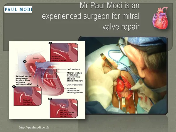
Mitral Incompetence • Valve does not close properly. • Blood flows back into the left atrium. • Volume overload of left ventricle. • Left ventricular failure.
Aetiology • Rheumatic Fever. • Endocarditis • Barlow's syndrome. (Floppy valve) • Ischemia. • Congenital. • Cardiomyopathy.
Carpentier classification • Type 1- Normal leaflet movement, annular dilatation. (cardiomyopathy) • Type 2- Increased leaflet movement, prolapsing segments. (Barlow's) • Type 3a- Restricted leaflet movement. ( Rheumatic) • Type 3b- Ischaemic leaflet retraction
Surgery • General anaesthesia. • TEE on board. • Cardio-pulmonary bypass. • Cell saver. • Repair before replace.
Type 2 – Valve prolapse • To much thickened leaflet. • Stretched out chordae. • Elongated papillary muscles. • Leaflet prolaps.
Type 3 b- Ischaemic incompetence • Valve dysfunction because of impaired coronary blood flow. • Posterior leaflet retraction. (P3 area) • Needs to be fixed > moderate incompetence. • Remodelling annuloplasty.
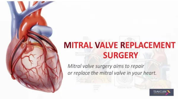
Mitral valve replacement • Native valve removed. • Mechanical or Tissue prosthesis.
Mechanical mitral valve replacement • Surgical mortality 2% - 4% • Bleeding risk 1%/year • Thrombo-embolism 1%/year • Endocarditis 0.1%/year