Download
1 / 41
470 likes | 1.34k Views
MORPHOLOGY OF THE MITRAL VALVE. M. Kuduvalli. ELEMENTS OF MITRAL VALVE APPARATUS. Annulus Leaflets Subvalvar apparatus - Chordae tendinae - Papillary muscles. MITRAL ANNULUS.

E N D
MORPHOLOGY OF THE MITRAL VALVE M. Kuduvalli
ELEMENTS OF MITRAL VALVE APPARATUS • Annulus • Leaflets • Subvalvar apparatus - Chordae tendinae - Papillary muscles
MITRAL ANNULUS • Zone of junction which serves as attachment to the muscular fibres of the atrium, ventricle, and attachment of the mitral valve • Attached to two fibrous trigones -The right fibrous trigone which forms a dense junction between the mitral, tricuspid and aortic (non-coronary cusp) annuli, and the membranous septum -The left fibrous trigone which lies between the aortic (left cusp) and the mitral annuli • Between the two trigones, the mitral valve is in continuity with the aortic wall and there is no fibrous mitral annulus in this region
SPATIAL RELATIONSHIP BETWEEN MITRAL, AORTIC AND TRICUSPID VALVES
MITRAL ANNULUS • Mitral annulus is a dynamic structure • Has a sphincter like function, effectively decreasing the valve area by about a quarter during systole • This is secondary to contraction and relaxation of the basoconstrictor muscles (bulbospiral and sinospiral) • Dilatation of the annulus occurs posteriorly
MITRAL LEAFLETS • Form a continuous veil attached to the circumference of the mitral annulus • Free edge hangs into the LV, and is split by indentations • Two well defined and constant indentations: - Anterolateral commissure - Posteromedial commissure
MITAL LEAFLETS • Commissural areas (identified by presence of commissural chordae) divide the continuous mitral veil into two leaflets: - Anterior (aortic) leaflet - Posterior (mural) leaflet
MITRAL LEAFLETS • Covered with endocardium • Distinct ridge on atrial side which - defines line of leaflet closure - separates leaflets into two zones - rough zone distal to the ridge (represents surface of coaptation) - clear zone proximal to the ridge
ANTERIOR MITRAL LEAFLET • Semicircular or triangular • Attached to around 3/8th of circumference of the mitral annulus • Has common attachment to the cardiac skeleton with - left coronary cusp of aortic valve - half of non-coronary cusp
ANTERIOR MITRAL LEAFLET • Rough zone receives the chordae tendinae • Forms boundary dividing the outflow and inflow tracts of the left ventricle.
ANTERIOR MITRAL LEAFLET • Direct continuity between AML and the aortic wall • Gap between aortic and mitral valves is filled with an inter-valvular septum. Fibrous mitral annulus is absent here 1.Intervalvular septum 2. AML 3. PML
POSTERIOR MITRAL LEAFLET • Quadrangular in shape • Attached to around 5/8th of the circumference of the mitral annulus • Margin has two indentations, forming three scallops: - Anterolateral - Middle - Posteromedial • Cleft chordae insert into these indentations
POSTERIOR MITRAL LEAFLET • Additional third zone, k/a basal zone, which is between the clear zone and the annulus. It receives insertion of the basal chordae • Basal zone is most obvious in the middle scallop since the majority of basal chordae insert here
SUBVALVAR APPARATUSPAPILLARY MUSCLES • Two groups of LV papillary muscles - Anterolateral - Posteromedial • Each group supplies chordae to their respective halves of both leaflets • Arise from the anterior and posterior walls of the left ventricle respectively
SUBVALVAR APPARATUSPAPILLARY MUSCLES • May have one or more bellies each. Anterolateral usually has one • Tip points towards the respective commissure
SUBVALVAR APPARATUSCHORDAE TENDINAE • Fibrous strings that originate from tiny nipples on the apical portion of the two papillary muscles • Majority have branching pattern, either soon after their origin from the papillary muscles, or just before their insertion into the leaflets
COMMISSURAL CHORDAE • Two in number, one for each commissure, with similar names • Arise as a main stem which branches radially to insert into the free margins of the commissural regions • Their attachment defines the extent of the commissural areas
CHORDAE OF THE A.M.L • Typically splits into 3 cords soon after its origin from the papillary muscles
MAIN CHORDAE OF THE A.M.L. • Two in number, one from each papillary muscle • Inserted at 4-5 O’clock posteromedially and 7-8 O’clock anterolaterally
OTHER CHORDAE OF THE A.M.L. • Paramedial chordae - Insert near the middle of the free edge • Paracommissural chordae - Insert between the main chordae and the commissural chordae
CHORDAE OF THE P.M.L. • Basal chordae - Unique to the PML - Arise directly as single strands from the left ventricular free wall or from the small trabeculum carnae • Rough zone chordae tendinae - Similar to AML chordae, but shorter and thinner • Cleft chordae - Insert into indentations on the PML
BLOOD SUPPLY OF THE MITRAL VALVE • Mitral leaflets and chordae are avascular • Papillary muscle supply - Anterolateral supplied by LAD and in addition, by the Diagonal or an OM from the Circumflex - Posterolateral variably supplied by branches of either the Lt. Circumflex or the RCA
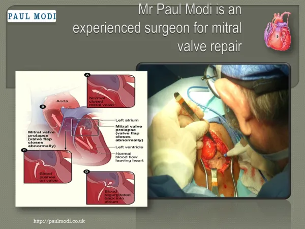
TYPES OF MITRAL VALVE PATHOLOGY • Type I: Normal leaflet motion - Annular dilatation - Leaflet perforation • Type II: Leaflet prolapse - Chordal rupture - Chordal elongation - Papillary muscle rupture - Papillary muscle elongation • Type III: Restricted leaflet motion - Restricted opening: Commissural fusion, leaflet and chordal thickening - Restricted closure: Excess tension on chordae during systole
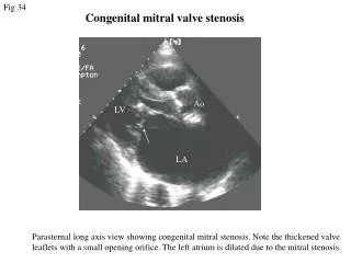
RHEUMATIC MITRALVALVE MORPHOLOGY • Can manifest as - Stenosis - Regurgitation - Mixed • Three primary pathological processes - Leaflet thickening - Chordal thickening, shortening and fusion - Coaptation of the edges of the leaflets, especially near the commissures
RHEUMATIC MITRALVALVE MORPHOLOGY • Leaflet thickening can progress to: - Calcification, first of leaflet, and then peri-annular - Retraction, leading to combined stenosis and regurgitation • Subvalvar apparatus involvment may lead to different degrees of subvalvar fusion
ISCHEMIC MITRAL VALVE DISEASE • Due to a combination of left ventricular wall akinesia or dyskinesia and ischemia of the papillary muscle itself, affecting the integrity of the subvalvar apparatus • Papillary muscle necrosis can lead to rupture either at its attachment at the base to the LV wall or at its tip near the chordal attachments • Leaflets and chordae are avascular structures, and are not directly involved in ischemic MR
MYXOMATOUS DEGENERATION MORPHOLOGY • Chordal elongation and rupture • Thickening of mitral leaflets • Redundancy of mitral leaflets, billowing into the left atrium in systole • Degeneration and abnormal collagen synthesis in the region close to the chordal attachments
INFECTIVE ENDOCARDITIS OF MITRAL VALVE • Leaflet involvement, with vegetation formation and subsequent destruction of the leaflet • Thickening and healing around chronic leaflet perforations • Annular and periannular abscesses, subsequently involving the aortic valve • Chordal detachment due to destruction of leaflet edges • Rupture of chordae and papillary muscles due to their primary involvement
OTHER DISEASES INVOLVING MITRAL VALVE • Marfan’s and Ehler-Danlos syndromes - Annular dilatation - Chordal elongation • Idiopathic calcification of the mitral annulus - particularly in the posterior area, with calcification extending into the LA - seen more frequently in elderly women
OTHER DISEASES INVOLVING MITRAL VALVE • HOCM – associated with MR - Distortion of the AML from contact with the hypertrophic IVS during systolic anterior motion of the AML - Dilatation of the LV and the annulus in long standing HOCM