Download
1 / 19
210 likes | 1.07k Views
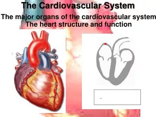
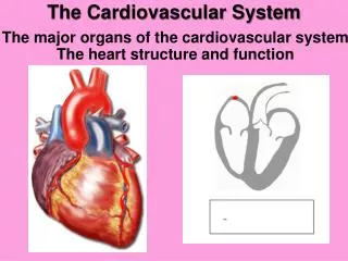
Interior & Conductive System of the Heart. The Heart The walls of the heart are composed of cardiac muscle. Out inward are: epicardium, myocardium endocardium. The Heart Is slightly larger than a clenched fist , working as double self-adjusting muscular pump.

E N D
The Heart The walls of the heart are composed of cardiac muscle. Out inward are: epicardium, myocardium endocardium
The Heart Is slightly larger than a clenched fist, working as double self-adjusting muscular pump. a. Human heart has four chambers 2 Atria 2 Ventricles b. The atria lie above & are separated from the ventricles by the atrioventricular groove/septum c. The atria are separated from each other by interatrial groove & ventricles by interventricular groove septum Left & Right
Interior of right atrium • Sulcus terminalis • a vertical groove on the outer side of heart at the junction b/w right atrium & auricle • d.Crista terminalis • inside the forms a ridge • c. Smooth posterior part main part Sinus Vinosus • d. Rough anterior part • musculi pactinati or pectinate muscles Crista terminalis
Openings into the Right Atrium • SVC • IVC • Coronary sinus • Right atrioventricular • valve • Many small veins Fetal remnants • Fossa ovalis • Anulus ovalis • Rudimentary valve of IVC
Interior of right ventricle • Is a triangular chamber • Upper part becomes funnel shaped • Infudubulum • C.Trabeculae carneae projecting ridges from the ventricular walls • composed of 3 types • i) papillary muscle attached with ventricular walls and apices are connected by fibrous chords • (chordae tendineae) to cusps of the tricuspid valve
ii) Attached two the ventricular walls without the middle part e.g. moderator band iii) Simply the prominent ridges.
Openings/valves in the right ventricle • Tricuspid valve • Anterior, Septal, & inferior cusps • the bases of cusps are attached to fibrous ring • of the skeleton of the heart • free tips thru Chordae tendineae to the • papillary muscles. During the ventricular • contraction prevent the regurgitation of blood • b. Pulmonary valve • Guards the pulmonary orifice • consists of 3 semilunar cusps
Right semilunar cusps Anterior semilunar cusps Left semilunar cusps
Interior of left atrium • The interior of the left atrium is smooth • Left auricle possess ridges
Openings into the Left Atrium • Four Pulmonary veins • Left atrioventricular orifice • guarded by bicuspid Mitral valve
Interior of Left Ventricle • The cavity s circular • Intraventricular BP is 6 times higher than RV • Well-developed trabeculae carneae, papillary muscles but no moderate bands like right ventricle • Walls are 3 times thicker • than right ventricle
Openings into the Left Ventricle • Bicuspid atrioventricular (Mitral) valve • Consists of two cusps (ant & post) • Similar to tricuspid valve • Tricuspid Aortic valve • Similar to the pulmonary valve • with right, left & posterior cusps • behind each cusp aortic wall bulges
Skeleton of the heart Consists of fibrous rings That surround the atrioventricular, Pulmonary & aortic orifices The fibrous rings support the bases of the cusps & prevent the valves from stretching & becoming incompetent
Conductive system of the heart Sinoatrial (SA) Node Located in the wall right atrium near the opening of the SVC Internodal Conduction Paths Atrioventricular (AV) Node Located in the atrial septum near the coronary sinus Atrioventricular Bundle Bundle of HIS Connects the myocardium of atria & ventricles Left & Right bundle branches Purkinje fiber