Download
1 / 54
550 likes | 575 Views
University Hospitals Case Medical Center Department of Radiology. Med Students Lecture Abdomen. The Gas Pattern – the crux of plain film in the abdomen. The gas pattern. Source swallowed air (majority) bacterial fermentation from food Normal Gas Pattern. The gas pattern.

E N D
University Hospitals Case Medical Center Department of Radiology Med Students LectureAbdomen
The gas pattern • Source • swallowed air (majority) • bacterial fermentation from food • Normal Gas Pattern
The gas pattern • Normal - Small bowel versus Large bowel
SMALL BOWEL LARGE BOWEL
The (abnormal) gas pattern • Localized Ileus • Focal irritation of loop(s) of bowel – adjacent inflammation • Causes – cholecystitis, pancreatitis, appendicitis, diverticulitis • Adynamic Ileus • Entire bowel is aperistaltic or hypoperistaltic • Cause - Postoperative Ileus – abdominal surgery • Mechanical Small Bowel Obstruction • Causes • Postsurgical Adhesions – most common • Malignancy – gastric, colon, ovarian • Hernia – inguinal • Gallstone ileus • Intussusception – Ileocolic (most common) • Inflammatory bowel disease
The (abnormal) gas pattern • Localized Ileus - Signs • 1 or 2 persistently dilated loops of small bowel (>3cm) • Air-fluid levels • Usually gas in the rectum and/or sigmoid colon This patient had underlying acute pancreatitis
The (abnormal) gas pattern • Adynamic Ileus (signs) • Diffuse small and large bowel dilitation • Air-fluid levels • Usually gas in the rectum and/or sigmoid colon Post-op Day 1
The (abnormal) gas pattern • Mechanical Small Bowel Obstruction (signs) • Multiple dilated loops of small bowel (step-ladder) • Numerous Air-Fluid Levels • Decompressed distal small bowel, No gas in rectum
The (abnormal) gas pattern • Mechanical Small Bowel Obstruction (signs)
The (abnormal) gas pattern • Mechanical Small Bowel Obstruction - adhesions
Using dilated loops of bowel for diagnostics • The location of dilated loops of bowel can be a sign of underlying pathology
Extraluminal air • Locations • Intrapeitoneal (pneumoperitoneum) • Retroperitoneal • Air in the bowel wall (pneumatosisintestinalis) • Air in the biliary tree (pneumobilia)
Extraluminal air • Intrapeitoneal (pneumoperitoneum) • Causes - rupture of air-containing structure • Perforated peptic ulcer • Trauma • Perforated diverticulitis/appendicitis • Perforation of carcinoma • Post-operative (5-7 days) • Signs • Crescenticlucency beneath the diaphram • Rigler’s Sign – Air on both sides of the bowel wall • Football Sign - Visualization of falciform ligament
Extraluminal air • Intraperitoneal Air (Pneumoperitoneum) • Crescentic lucency beneath the diaphram
Extraluminal air • Intraperitoneal Air (Pneumoperitoneum) • Rigler’s Sign – Air on both sides of the bowel wall
Extraluminal air • Intraperitoneal Air (Pneumoperitoneum) • Football sign - Visualization of falciform ligament
Extraluminal air • Retroperitoneal • Causes • Rupture of bowel (e.g. ruptured appendicitis or UC) • Trauma – blunt or penetrating • Iatrogenic manipulation – surgery or colonoscopy • Foreign body – causing perforation • Gas-producing infection – perforated diverticulitis
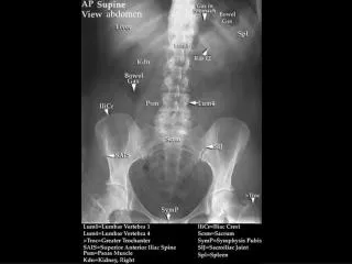
Extraluminal air • Retroperitoneal - Signs • Streaky, linear appearance outlining extraperitoneal structures (e.g. psoas muscle, kidneys, bladder, aorta, IVC) • Mottled, blotchy appearance – remain in a fixed position
Extraluminal air • Air in the bowel wall (pneumatosisintestinalis) • Causes • Primary form – pneumatosiscystoides • Left colon – cyst-like collections of air – submucosa/serosa • Secondary form – obstructive and necrotizing disease • Chronic obstructive pulmonary disease • Necrosis of bowel • Necrotizing enterocolitis – MOST COMMON CAUSE IN infants • Ischemic bowel – MOST COMMON CAUSE IN adults • Obstructing lesions of the bowel • Hirshsprung’s disease/Pyloric stenosis – children • Obstructing carcinoma - adults
Extraluminal air • Air in the bowel wall (pneumatosis intestinalis) • Signs • Linear radiolucency paralleling the contour of air in the adjacent bowel lumen
Extraluminal air • Air in the bowel wall (pneumatosis intestinalis) • Signs • Mottled appearance • resembles air mixed with fecal material
Extraluminal air • Air in the biliary tree (pneumobilia) • Causes • Incompetent sphincter of Oddi • Prior Sphincterotomy • Prior surgery – reimplantation of the common bile duct • Gallstone ileus • Gallstone erodes through the wall of the gallbladder into the duodenum – forms a fistula between the bowel and biliary system • Gas-forming pyogenic cholangitis
Extraluminal air • Air in the biliary tree (pneumobilia) • Signs • Tube-like, branching lucencies in the right upper quadrant – overlying the liver • Air in the wall of the gallbladder
Types of procedures done by radiologists in the abdomen • Cryoablations – treatment for cancers of the liver and kidney • Biopsies of every organ • Intravascular stenting • Treatment of aortic aneurysms and arterial atherosclerosis • IVC filters for PE prevention • Treatment of renal artery stenosis for hypertension • GI bleed management • Drainages of abscess • Percutaneous nephrostomy placement • Percutaneous cholecystostomy placements • PEG tube placement • TIPS And many many more!!
GI Bleeds • IR plays a big role in the management of significant bleeds • First step in management depends on types of bleed and patient status • Intervention is rarely step 1
GI Bleeds • Types of GI bleeds • Upper GI Bleed • Presents with melena (usually), hematemesis • Major causes?? (ask the students):
GI Bleeds • Types of GI bleeds • Upper GI Bleed • Presents with melena (usually), hematemesis • Major causes?? (ask the students): • Ulcers • Gastritis • Esophagitis • Esophageal varices
GI Bleeds • Types of GI bleeds • Lower GI Bleed • Presents with hematochezia • Major causes?? (ask the students):
GI Bleeds • Types of GI bleeds • Lower GI Bleed • Presents with melena, hematemesis • Major causes?? (ask the students): • Diverticulosis • Angiodysplasia • Rectal problems – hemorrhoids, fissures
GI Bleed • Management – • Is the patient stable? • Yes ---> can manage conservatively first, then pursue interventional techniques • No SURGICAL or ENDOSCOPIC management to identify site of bleed • Where is the bleed? • Bright red blood start with rectal exam to rule out hemorrhoids (very common) • Check history and diet – certain meds/vitamins can make stool look red • After history and physical, proceed to imaging if still needed
GI Bleeds • Choosing imaging relies on frequency of the bleed • High volume and frequency of bloody bowel movements • Proceed to contrast enhanced CT angiogram • Low frequency or volume • Proceed to nuclear tagged red blood scan **generally, imaging is required prior to IR embolization to help localize the source and plan the procedure exceptions – if source is obvious from history or patient has contraindications to contrast and cant get nuclear imaging
GI Bleeds • Management: • If patient is stable, always start with fluids and/or pRBCs • Majority of bleeds stop on their own with conservative management • If persistent bleeding despite conservative management • IR guided embolization – • Femoral arterial approach angiograms of the celiac, SMA, and IMA are done to localize the site Bleeding rectal varices seen on IMA angiogram
GI Bleeds • IR embolization can be done with • Coils – permanent • Gelfoam – temporary • Decision on what to use depends on patient stability, degree of bleeding, and risk of necrosis based on vascular territories involved with embolization
Imaging modalities in Abdomen • Abdominal xray • Great for looking for obstructions and gas pattern • Bad for evaluating individual organ pathology • Fluoroscopy • Best for evaluating real-time pathology of the GI and GU tracts • CT • Best screening for evaluating pathology of individuals organs • MRI • Best for better characterizing findings on CT • Ultrasound • Best for dynamic real-time imaging and evaluation of vasculature
Appropriate Imaging • Abdominal xray is usually the best imaging to start with • This can give an idea of the bowel pattern • Can be done portably – better idea for ICU patients • CT • Non-radiologists should know when to order contrast • Types of contrast: • IV – best for highlighting pathology within all organs • Oral – best for differentiating the GI tract from other pathology OR evaluating for obstructions • Rectal – best for evaluating trauma to the colon
Common diagnoses • Abdominal distention – start with an xray for the bowel pattern – evaluate for gastric distention, SBO or ileus • If the xray is normal, CT is not very useful • A combination of xrays and physical exam can help determine the presence of ascites • Trauma – start with CT with IV contrast • Jaundice – depends on the likely cause – use liver function tests to determine the likely cause • Ultrasound or CT/MR for liver • Ultrasound for biliary pathology • CT or MRCP for pancreas
Common diagnoses • Hematuria – need to rule out bladder or renal CA • CT with IV contrast is the best place to start • MR if there is known pathology • Flank pain – need to look for stones • CT WITHOUT contrast – IV will obscure stones
Common diagnoses • Dysphagia – often subjective, but can be caused by obstructions, stenoses, or muscular problems • FIRST: determine the type of dysphagia • Solids versus liquids • Upper esophagus, swallowing problems versus Mid or Lower Chest dysphagia • THEN determine the type of study • Modified barium swallow – evaluates upper esophageal function including the hypopharnyx and oropharynx • Performed by speech pathology • Barium esophogram – evaluates esophageal function in the mid/lower esophagus – good for postoperative evaluation or mid-sternal dysphagia • Performed by radiologist
Common diagnoses • Left lower quadrant pain – likely diverticulitis • CT with IV contrast – NOT for the primary diagnosis • Looking for complications such as abcess • Right lower quadrant pain – likely appendicitis • CT with IV contrast – again, looking for complications • Right upper quadrant pain – likely gallbladder pathology • Start with ultrasound - CT is not as sensitive • Proceed to CT if ultrasound is negative or equivocal • Left upper quadrant pain – pancreatitis or ulcer disease • Imaging is less useful • CT findings of pancreatitis lag behind lipase elevation – no use of imaging for diagnosis • CT is used to detect complications if patients are not improving – NOT needed for primary diagnosis
Special imaging populations • Right and left lower quadrant pains in: • Children • Pathologies are different • RLQ – appendicitis • LLQ – intusseception • Start with ultrasound – CT has too much radiation and lower sensitivity in peds • Women • Different pathologies – ovarian pathologies are also a consideration • Determining which to start with, ultrasound or CT, relies on good clinical skills (history taking and physical exam)
Special imaging populations • Infants • Most common abdominal complaints are: • Projectile vomiting – think pyloric stenosis • Ultrasound is first step • Bilious emesis – think malrotation • Upper GI fluoroscopic study is the first step • Abdominal pain – think intussception • Ultrasound is the first step