Download
1 / 39
1.03k likes | 2.85k Views
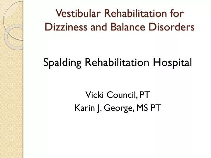
Vestibular Rehabilitation for Dizziness and Balance Disorders. Spalding Rehabilitation Hospital Vicki Council, PT Karin J. George, MS PT. Life is Good. Objectives. Be familiar with the anatomy and physiology of the vestibular system

E N D
Vestibular Rehabilitation for Dizziness and Balance Disorders Spalding Rehabilitation Hospital Vicki Council, PT Karin J. George, MS PT
Objectives • Be familiar with the anatomy and physiology of the vestibular system • Have a basic understanding of the assessments for vertigo and balance • Be familiar with the treatment strategies to address vestibular and balance disorders
Vestibular Rehabilitation • Evaluation to discover deficits and details regarding symptoms • Develop program with goal of retraining the brain to recognize and process signals from the vestibular system in coordination with the visual and proprioceptive systems.
A Need for Intervention • ~4% (8 million) of American adults report having a chronic balance problem. An additional 1.1% (2.4 million) report chronic problems with dizziness alone. • Overall, the cost of medical care for patients with balance disorders exceeds $1 billion/year in the United States. • Though BPPV is seen by most as the most common vestibular disorder, only ~8% receive effective tx.
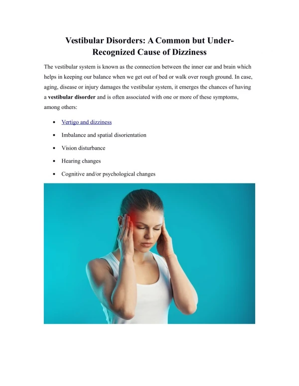
Definitions • Dizziness • Light headedness • Feeling faint • Unsteady • Dysequilibrium
Definitions • Vertigo • A specific spinning sensation • An illusion of motion • The feeling that you or your environment is moving
Definitions • Imbalance • Stumbling, difficulty walking straight or turning a corner • Clumsiness or difficulty with coordination • Tendency to fall
Balance Systems • Sensory • Visual - eyes orient • Vestibular - inner ear system that registers the head and movement in space • 3 components • Gaze stabilization • Postural Control • Perception of movement • Proprioceptive - involves the joint and muscle receptors that let us know where we are in space
Balance Systems • Motor • Strength • Coordination • Cardiorespiratory/endurance • ROM • Reaction time
Anatomy and Physiology of the Inner Ear • Bony Labyrinth: Houses the membranous labyrinth. Filled with perilymph fluid (similar to CSF). • Membranous Labyrinth: Consists of the otolith organs and semi-circular canals. Filled with endolymph (similar to intracellular fluid).
Membranous Labyrinth • Otolith Organs • Utricle: Responsible for horizontal translation of the head and head tilt • Saccule: Responsible for vertical translation of the head. • Combined, they sense linear acceleration and static tilt of the head with respect to the gravitational axis. • Maculae: Sensory receptor for the otolith organs. A gelatinous matrix surrounds the hair cells. Otoconia are embedded on top of the maculae in the otolithic membrane..
Membranous Labyrinth • Semi-circular Canals (SSC) • Three fluid filled loops responsible for sensory input related to head velocity and angular acceleration. • Enables the VOR to generate eye movements to match head movements, resulting in clear vision during head movement. • Cristae: sensory structure for the SSC that sense angular movement • Cupula: gelatinous mass surrounding the hair cells of the cristae in the SSC.
Membranous Labyrinth • Hair Cells: • Endolymph fluid within the SSC and otolith organs move the hair cells according to head movement. • The direction of deflection of the hair cells tells the brain how the head is moving. Resting Vestibular Tone: Firing rate = 100 spikes/sec at rest, increases on ipsilateral side and decreases on contralateral side related to head movement.
Central Processing • Electrical activity generated from the inner ear travels to: • Vestibular nuclei in the brainstem • Cerebellum • Emetic center in the brainstem
Dizziness Diagnosis • Unilateral Peripheral Vestibular Loss • Nystagmus • Falling/loss of balance to affected side • Possible Cerebellar Dysfunction • True Vertigo • Bilateral Peripheral Vestibular Loss • Usually no vertigo • Oscillopsia • Ataxia • Imbalance
Dizziness Diagnosis • Related to specific health issues: • Vestibular Neuritis • Viral Labyrinthitis • CVA • Acoustic neuroma • Ototoxicity
Vestibular Pathologies Seen in Head Injury • Endolymphatic hydrops or post-traumatic Meniere’s disease • Vestibular migraine • Labyrinthine concussion • Perilymphatic fistula • Brainstem injury
Dizziness Diagnosis • Duration of Spells: • Seconds: BPPV Orthostatic Hypotension • Minutes: Migraines TIAs • Hours/Days: Meniere’s Disease Hydrops
ENT Testing • Hearing Test • Videonystagmography (VNG)- test of lateral semi-circular canal • Vestibular evoked myogenic potentials (VEMP)-test of otolithic function • Electrocochleography (ECoG)- test for endolymphatic hydrops (Meniere’s disease) • Platform Posturography- Assesses use of all sensor systems-visual, vestibular, somatosensory
Vestibular Evaluation • Subjective • Dizziness • What makes it better, worse, duration, frequency, intensity • Falls • Dizziness Handicap Index • History including test results/medications
Vestibular Evaluation • Objective • Vision and eye/head coordination • Eye movement range • Smooth pursuits • End point nystagmus • Gaze evoked nystagmus • Saccades • Skew eye deviation • Spontaneous nystagmus • VOR Cancellation • Visual/Vestibular Ocular Interaction Reflex
Vestibular Evaluation • Vertiginous Positions/movements: requires that the patient be placed in 16 positions monitoring level of dizziness, nystagmus and length of time of dizziness. • Hallpike assesses BPPV
Vestibular Evaluation • Strength • ROM • Joint position sense • Sensation • Gait
Vestibular Evaluation • Balance Tests • BERG • Dynamic Gait • Balance Evaluation-Systems Test (BEST) • Balance Master(sensory organization test)
Smart Balance Master Sensory Organization Test • Force plate system • Moving surround • Uses 3 senses for postural control-vestibular, visual, somatosensory. • Looks at the ability to differentiate the differences in accurate inputs, choose the most useful input, and perceive the correct position in space.
Red Flags • Sudden loss of hearing or fluctuation in hearing • Pressure and fullness to point of pain/discomfort • Fluid from ears • Severe ringing in ears • Other undiagnosed central signs
Medications • Anticholinergics – Antihistamines • Meclizine • Antivert • Phenergan • Benzodiazepines • Valium • Klonapin • Ativan
Vestibular Treatment • Central Compensation • Adaptive Plasticity • Central Sensory Substitution • Tonic Rebalancing • Habituation
Treatment • Symptom driven • Head/eye movements • Position progression • Support surface • Eyes open/closed • Environment • Manual Interventions • Canalith Repositioning (BPPV)
Vestibular Treatment • Rehab strategies: • Retraining • Habituation • Adaptation • Compensation
Vestibular Treatment • Motor Learning • HEP based on deficits • Education • Manual Interventions • Referrals to other providers
Time Frames • Simple • 3-4 sessions • Complex-involving multiple systems • 6-18 months • Patient compliance • 6 to 18 months
Team Role • Know general signs and symptoms • Gather information and refer patient to appropriate specialists • Repeat education to patient and family • VEDA (Vestibular Disorders Association)