Download
1 / 85
850 likes | 949 Views
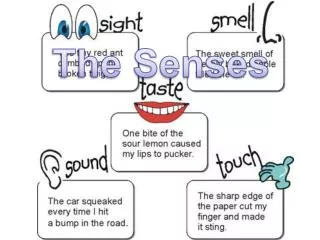
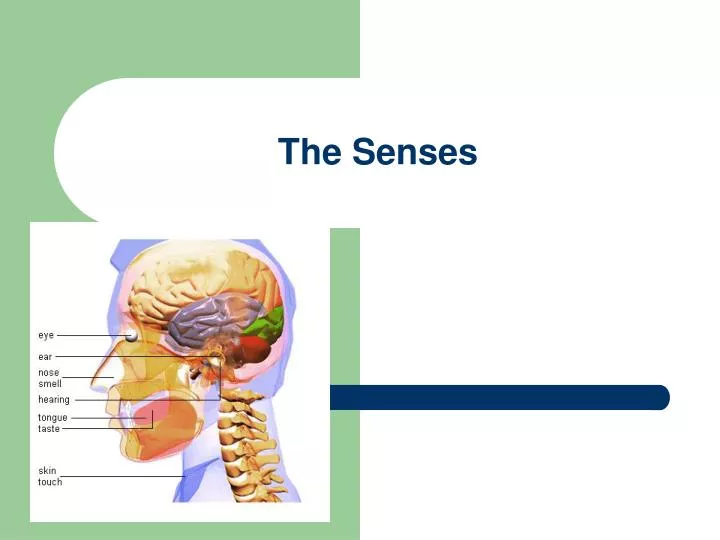
The Senses. Perception Behavior. Types of Receptors. Mechanoreceptors – stimulated by mechanical energy Chemoreceptors – detect solute concentration differences Electromagnetic receptors – detect forms of electromagnetic energy Thermoreceptors – respond to hot or cold

E N D
Types of Receptors • Mechanoreceptors – stimulated by mechanical energy • Chemoreceptors – detect solute concentration differences • Electromagnetic receptors – detect forms of electromagnetic energy • Thermoreceptors – respond to hot or cold • Pain receptors – naked dendrites in epidermis of skin
Touch • Sensory receptors in the skin receive the touch stimulus • Mechanoreceptors in human skin are in the form of naked dendrites • Prostaglandins intensify the pain by sensitizing the receptors
Extrinsic Eye Muscles Six straplike extrinsic eye muscles Enable the eye to follow moving objects Maintain the shape of the eyeball Four rectus muscles originate from the annular ring Two oblique muscles move the eye in the vertical plane Sight
The retina at the back of the eye light receptors and sensory neurons. Rods= adapt vision in dim light. Cones= detect color. Tissue comes together to form the otic nerve which carries impulses directly to the brain. Sight
Fibrous Tunic • Forms the outermost coat of the eye and is composed of: • Opaque sclera (posteriorly) • Clear cornea (anteriorly) • The sclera protects the eye and anchors extrinsic muscles • The cornea lets light enter the eye
Vascular Tunic: Ciliary Body • A thickened ring of tissue surrounding the lens • Composed of smooth muscle bundles (ciliary muscles) • Anchors the suspensory ligament that holds the lens in place
The colored part of the eye Pupil – central opening of the iris Regulates the amount of light entering the eye during: Close vision and bright light – pupils constrict Distant vision and dim light – pupils dilate Changes in emotional state – pupils dilate when the subject matter is appealing or requires problem-solving skills Vascular Tunic: Iris
A delicate two-layered membrane Pigmented layer – the outer layer that absorbs light and prevents its scattering Neural layer, which contains: Photoreceptors that transduce light energy Bipolar cells and ganglion cells Amacrine and horizontal cells Sensory Tunic: Retina
Ganglion cell axons: Run along the inner surface of the retina Leave the eye as the optic nerve The optic disc: Is the site where the optic nerve leaves the eye Lacks photoreceptors (the blind spot) The Retina: Ganglion Cells and the Optic Disc
The Retina: Photoreceptors • Rods: • Respond to dim light • Are used for peripheral vision • Cones: • Respond to bright light • Have high-acuity color vision • Are found in the macula lutea • Are concentrated in the fovea centralis
What sort of neuro-transmitters must be released from the rod cell to neurons in the dark?
Why are you temporarily blinded when you enter a dark movie theatre on a sunny day?
Visual integration: • Receptive fields feed information to one ganglion cell • Larger receptive fields result in a less sharp image • Ganglion cells of fovea have small receptive fields
Blood Supply to the Retina • The neural retina receives its blood supply from two sources • The outer third receives its blood from the choroid • The inner two-thirds is served by the central artery and vein • Small vessels radiate out from the optic disc and can be seen with an ophthalmoscope
Inner Chambers and Fluids • The lens separates the internal eye into anterior and posterior segments • The posterior segment is filled with a clear gel called vitreous humor that: • Transmits light • Supports the posterior surface of the lens • Holds the neural retina firmly against the pigmented layer • Contributes to intraocular pressure
Composed of two chambers Anterior – between the cornea and the iris Posterior – between the iris and the lens Aqueous humor A plasmalike fluid that fills the anterior segment Drains via the canal of Schlemm Supports, nourishes, and removes wastes Anterior Segment
Lens • A biconvex, transparent, flexible, avascular structure that: • Allows precise focusing of light onto the retina • Is composed of epithelium and lens fibers • Lens epithelium – anterior cells that differentiate into lens fibers • Lens fibers – cells filled with the transparent protein crystallin • With age, the lens becomes more compact and dense and loses its elasticity
Electromagnetic radiation – all energy waves from short gamma rays to long radio waves Our eyes respond to a small portion of this spectrum called thevisible spectrum Different cones in the retina respond to different wavelengths of the visible spectrum Light
When light passes from one transparent medium to another its speed changes and it refracts (bends) Light passing through a convex lens (as in the eye) is bent so that the rays converge to a focal point When a convex lens forms an image, the image is upside down and reversed right to left Refraction and Lenses
Focusing Light on the Retina • Pathway of light entering the eye: cornea, aqueous humor, lens, vitreous humor, and the neural layer of the retina to the photoreceptors • Light is refracted: • At the cornea • Entering the lens • Leaving the lens • The lens curvature and shape allow for fine focusing of an image
Light from a distance needs little adjustment for proper focusing Far point of vision – the distance beyond which the lens does not need to change shape to focus (20 ft.) Focusing for Distant Vision
Problems of Refraction • Emmetropic eye – normal eye with light focused properly • Myopic eye (nearsighted) – the focal point is in front of the retina • Corrected with a concave lens • Hyperopic eye (farsighted) – the focal point is behind the retina • Corrected with a convex lens
Photoreception – process by which the eye detects light energy Rods and cones contain visual pigments (photopigments) Arranged in a stack of disklike infoldings of the plasma membrane that change shape as they absorb light Photoreception: Functional Anatomy of Photoreceptors
Functional characteristics Sensitive to dim light and best suited for night vision Absorb all wavelengths of visible light Perceived input is in gray tones only Sum of visual input from many rods feeds into a single ganglion cell Results in fuzzy and indistinct images Rods
Excitation of Cones • Visual pigments in cones are similar to rods (retinal + opsins) • There are three types of cones: blue, green, and red • Intermediate colors are perceived by activation of more than one type of cone • Method of excitation is similar to rods
Functional characteristics Need bright light for activation (have low sensitivity) Have pigments that furnish a vividly colored view Each cone synapses with a single ganglion cell Vision is detailed and has high resolution Cones
Eye and Associated Structures • 70% of all sensory receptors are in the eye • Most of the eye is protected by a cushion of fat and the bony orbit • Accessory structures include eyebrows, eyelids, conjunctiva, lacrimal apparatus, and extrinsic eye muscles
Eyebrows • Coarse hairs that overlie the supraorbital margins • Functions include: • Shading the eye • Preventing perspiration from reaching the eye • Orbicularis muscle – depresses the eyebrows • Corrugator muscles – move the eyebrows medially
Protect the eye anteriorly Palpebral fissure – separates eyelids Canthi – medial and lateral angles (commissures) Palpebrae (Eyelids)
Conjunctiva • Transparent membrane that: • Lines the eyelids as the palpebral conjunctiva • Covers the whites of the eyes as the ocular conjunctiva • Lubricates and protects the eye
Consists of the lacrimal gland and associated ducts Lacrimal glands secrete tears Tears Contain mucus, antibodies, and lysozyme Enter the eye via superolateral excretory ducts Exit the eye medially via the lacrimal punctum Drain into the nasolacrimal duct Lacrimal Apparatus
1. Cornea 2. Anterior Chamber 3. Pupil 4. Lens 5. vitreous Chamber 6. Retina (Contain: rods (shades) and cones (color) 7. Optic disk 8. brain Pathway of light (image) through eye:
Taste and Smell • Chemoreceptors sense chemicals in the environment • Olfactory receptors line nasal cavity • Taste receptors respond to specific stimuli (sugar/ salt) • Taste and smell are functionally similar: • Molecule dissolves in liquid to reach receptor • Head cold interferes with taste perception
Most of the 10,000 or so taste buds are found on the tongue Taste buds are found in papillae of the tongue mucosa Papillae come in three types: filiform, fungiform, and circumvallate Fungiform and circumvallate papillae contain taste buds Taste Buds
Taste Sensations • There are five basic taste sensations • Sweet – sugars, saccharin, alcohol, and some amino acids • Salt – metal ions • Sour – hydrogen ions • Bitter – alkaloids such as quinine and nicotine • Umami – elicited by the amino acid glutamate
Physiology of Taste • In order to be tasted, a chemical: • Must be dissolved in saliva • Must contact gustatory hairs • Binding of the food chemical: • Depolarizes the taste cell membrane, releasing neurotransmitter • Initiates a generator potential that elicits an action potential