Download
1 / 56
560 likes | 810 Views
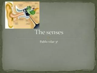
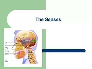
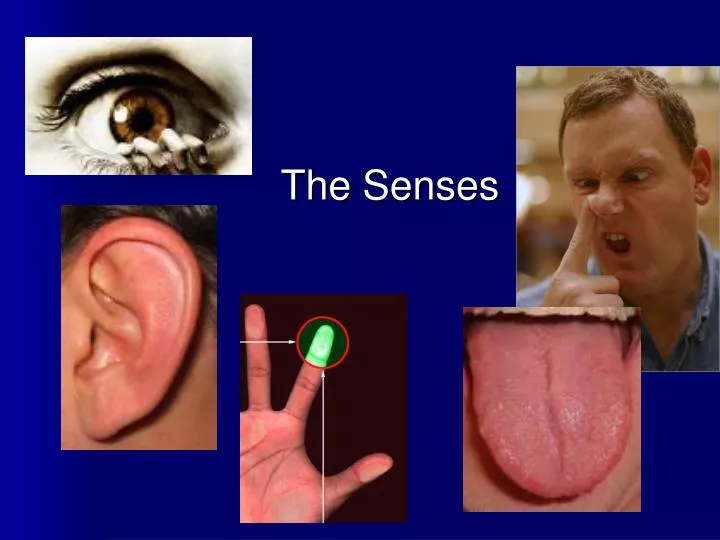
The Senses. The Senses. General senses of touch Temperature Pressure Pain Special senses Smell / Taste Sight Hearing / Equilibrium (balance). The Special Senses. Sight – The eye and Vision Smell / Taste – Chemical senses – Nose and Tongue Hearing / Equilibrium (balance) – The Ear.

E N D
The Senses • General senses of touch • Temperature • Pressure • Pain • Special senses • Smell / Taste • Sight • Hearing / Equilibrium (balance)
The Special Senses • Sight – The eye and Vision • Smell / Taste – Chemical senses – Nose and Tongue • Hearing / Equilibrium (balance) – The Ear
The Eye • Visual organ – the eye • 70% of all sensory receptors are in the eyes. • 40% of the cerebral cortex is involved in processing visual information. • Each eye has over a million nerve fibers. • Protection for the eye • Most of the eye is enclosed in a bony orbit, in other words your eye socket. • A cushion of fat surrounds most of the eye.
Accessory Structures of the Eye • Eyelids Designed to protect the eye, and keep moisture distributed over the surface of the eyeball. Slide 8.3a
Accessory Structures of the Eye • Eyelashes Acts as a dust and particle protector for the eye. Has modified sebacious glands produce an oily secretion to lubricate the eye. • Ciliary glands modified sweat glands between the eyelashes.
Tear ducts or the Lacrimal apparatus • Tears contain mucous, antibodies, (anti-bacterial) • keeps the surface of the eye moist. • Lacrimal gland – produces the tears. • Lacrimal sac – fluid empties into nasal cavity.
Eye Muscles • Muscles attach to the outer surface of the eye. • Produce eye movements.
Structure of the Eye The wall of the eye is composed of three tunics • Sclera & Cornea fibrous outside layer • 2. Choroid – middle layer • 3. Sensory tunic – (retina) inside layer.
1. The Fibrous Tunic • Sclera • Tough white connective tissue layer. • The “white of the eye” • Cornea • Transparent, central anterior portion. • Allows for light to pass through. • Repairs itself easily. • The only human tissue that can be transplanted without fear of rejection.
Choroid Layer • Blood-rich nutritive tunic • Pigment prevents light from scattering. • Modified interiorly into two structures. • Cilliary body – smooth muscle • Iris Pigmented layer that gives eye color Pupil – rounded opening in the iris
Sensory Tunic (Retina) • Contains receptor cells (photoreceptors) • Rods • Cones • Signals pass from photoreceptors and leave the retina toward the brain through the optic nerve
Neurons of the Retina and Vision • Cones – 3 types detect different colors • Densest in the center of the retina. • Fovea centralis – area of the retina with only cones. • Lack of one type = color blindness.
Neurons of the Retina and Vision • Rods • Most are found towards the edges of the retina • Allow dim light vision and peripheral vision • Perception is all in gray tones
The Iris • Visible colored part of the eye • Composed of smooth muscle • Pupil – the round, central opening that is a set of special muscles which acts to vary the amount of light entering the eye.
Lens • Biconvex crystal-like structure • Held in place by ligaments.
Lens – what it does • Light must be focused to a point on the retina for optimal vision • The eye is set for distance vision (over 20 ft away) • The lens must change shape to focus for closer objects Slide 8.16
Internal Eye Chamber Fluids • Vitreous humor • Keeps the eye from collapsing • Gel-like substance behind the lens • Lasts a lifetime and is not replaced • Aqueous humor • Similar to blood plasma • Watery fluid found in chamber between the lens and cornea • Provides nutrients for the lens and cornea
Vision • Each eye captures its own view and the two separate images are sent on to the brain for processing. • When the two images arrive simultaneously in the back of the brain, they are united into one picture. • The mind combines the two images by matching up the similarities and adding in the small differences. • The combined image is more than the sum of its parts. It is a three-dimensional stereo picture.
BLIND SPOT • The area on the retina where the optic nerve enters the eyeball. • This area has no photoreceptors and therefore no visual input. • The cortex appears to fill-in this missing information so we are not conscious of the blind spot. • No photoreceptor cells are at the optic disk, or blind spot. BLIND SPOT – little test Slide 8.16
The Eye - basic parts review http://www.bpei.med.miami.edu/site/disease/disease_anatomy.asp
Correcting the Eye • Nearsightedness = myopia • Focus of light in front of retina • Eyeball too long or lens too strong • Distant objects are blurry • Farsightedness = hyperopia • Focus of light beyond the retina • Short eyeball or lazy lens • Near objects are blurry. • Difficulty seeing clase objects = presbyopia • Inability of the lens to focus properly at close objects • Caused by the aging of the eye. • Special reading glasses needed.
Cataracts • The natural lens looses its transparency due to damage to its fibers over time. • Lens fibers are not replaced. • When the lens of the eye turns cloudy enough to impair vision, it is considered a cataract. • They are the main cause of blindness worldwide. • Most individuals over 60 years old develop some degree of cataract. • Treatment consists of a safe and precise surgical procedure.
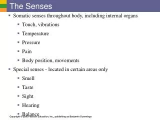
The Ear • Houses two senses • Hearing • Equilibrium (balance) • Receptors are mechanoreceptors, they react to sound waves.
Anatomy of the Ear • The ear is divided into three areas • Outer (external) ear • Middle ear • Inner ear
The External Ear Involved in hearing only • Structures of the external ear • Pinna (auricle) • External auditory canal
The External Auditory Canal • Narrow chamber in the temporal bone • Lined with skin • Ceruminous (wax) glands are present • Ends at the tympanic membrane or ear drum.
The Middle Ear or Tympanic Cavity • Air-filled cavity within the temporal bone • Only involved in the sense of hearing
The Middle Ear or Tympanic Cavity • Two tubes are associated with the inner ear • The opening from the auditory canal is covered by the tympanic membrane (Ear drum) • The auditory tube connecting the middle ear with the throat • Allows for equalizing pressure during yawning or swallowing • This tube is otherwise collapsed
Bones of the Tympanic Cavity • Three bones span the cavity (the smallest bones in our bodies!!) • Malleus (hammer) • Incus (anvil) • Stapes (stirrip)
Vibrations from eardrum move the malleus • These bones transfer sound to the inner ear. Slide 8.25b
Inner Ear or Bony Labyrinth • Includes sense organs for hearing and balance! • Filled with a fluid called perilymph Slide 8.26a
Inner Ear or Bony Labyrinth • A maze of bony chambers within the temporal bone • Cochlea • Vestibule • Semicircular canals
Hearing • Located within the cochlea • Receptors = hair cells a membrane on it’s inner surface. • Cochlear nerve attached to hair cells transmits nerve impulses to auditory cortex on temporal lobe of the brain.
Equilibrium – Balance/Orientation • Receptor cells are in two structures: • Vestibule • Semicircular canals
Equilibrium Equilibrium has two functional parts • Static equilibrium – sense of gravity at rest. Ability to stay still in one place. • Dynamic equilibrium – angular and rotary head movements. Keeping a sense of where you are at all times Think of a snowboarder doing a flip and being able to land on their feet. Figure 8.16a, b
Equilibrium This balance is achieved by vestibular nerve endings in side the Vestibule and the Semicircular canals, sensing the subtle changes in the fluid (endolymph) inside these structures. Figure 8.16a, b
Chemical Senses – Taste and Smell • Both senses use chemoreceptors • Stimulated by chemicals in solution. • Taste has four types of receptors. • Smell can differentiate a very large range of chemicals. **Both senses complement each other and respond to many of the same stimuli**
Olfaction – The Sense of Smell • Olfactory receptors are in the roof of the nasal cavity. • Neurons with long cilia • Chemicals must be dissolved in mucus for detection • Impulses are transmitted via the olfactory nerve • Interpretation of smells is made in the cortex of the Brain
Taste • Taste buds house the receptor organs • Location of taste buds • Most are on the tongue Slide 8.37