Download
1 / 69
710 likes | 1.12k Views
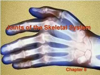
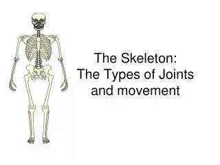
The Joints of the Skeleton System. Primarily Synovial Joints. JOINTS. Joints or articulations are functional junction between bones…or the site where two bones meet.

E N D
The Joints of the Skeleton System Primarily Synovial Joints
JOINTS • Joints or articulations are functional junction between bones…or the site where two bones meet. • They bind parts of the skeletal system, make possible bone growth, permit parts of the skeleton to change shape during childbirth, and enable the body to move in response to skeletal muscle contractions.
Joints can also be grouped according to the degree of movement possible at the bony junction. Synarthrotic – Immovable joints Amphiarthrotic- Slightly moveable Diarthrotic- Freely movable. JOINTS
Joints vary considerably in structure and function However, they can be classified by the type of tissue that binds the bones at each junction. Three general groups are: Fibrous joints Cartilaginous Joints Synovial Joints JOINTS
Fibrous Joints • Fibrous Joints- are so named because of the fibrous tissue that holds them together • In a fibrous joint there is no joint cavity present. • Most fibrous joints are immovable. • They lie between bones that closely contact one another.
Fibrous Joints • Three types of fibrous joints: • Syndesmosis-Distal end of the tibia and fibula • Suture- only between flat bones in the skull • Gomphosis- root of a tooth to the jawbone
Cartilaginous Joints • Cartilaginous Joints- Hyaline cartilage or fibrocartilage connect the bones of this joint. • Two Types are: • Synchondrosis (the articulation between the first rib and the manubrum) • Symphysis (the smyphiysis pubis in the pelvis)
SYNOVIAL JOINTS • Most joints of the skeletal system are synovial joints, and because they allow free movement, they are diarthrotic. • These joints are more complex structurally than fibrous or cartilaginous joints.
SYNOVIAL JOINTS • Synovial Joint have five distinguishing features. • Articular Cartilage is present at the ends of the opposing bones • Joint Cavity (synovial) is present. This is really just potential space that contains fluid. • Articular capsule (aka joint capsule). This encloses the joint cavity. • Synovial Fluid- is present and occupies the space within the joint capsule. • Reinforcing Ligaments – Synovial joints are reinforced and strengthened by a number of bandlike ligaments.
Bursae and Tendon Sheaths • Not strictly associated with synovial joints but are often found associated with them. • Bursae are flattened fibrous sacs lined with synovial membrane and containing a thin film of synovial fluid. • Tendon Sheath is essentially an elongated bursa that wraps completely around a tendon subjected to friction.
The Ball and Socket joint consists of a bone with a globular or slightly egg- shaped head the articulates with the cup-shaped cavity of another. Such joint allows a wider range of motion than does any other kind. Permitting movements in all planes, as well as rotation movement around a central axis. Ball and Socket
Ball and Socket • Examples: Hip and Shoulder contain joints of this type.
Condyloid joint, the oval condyle of one bone fits into the elliptical cavity of another bones. This type of joints permits a variety of movements in different planes; rotational movement, however,is NOT possible. Condyloid Joint
Condyloid Joint • Condyloid Joint exists between the metacarpals and the phalanges. • And the radiocarpal (wrist) joints.
The articulating surfaces of gliding joints are nearly flat or slightly curved. These joints allow sliding or back-and-forth motion and twisting movements. GLIDING JOINTS(aka Plane Joints)
GLIDING JOINTS(aka Plane Joints) • Most of the joints within the wrist and ankle. • The articular processes of adjacent vertebrae. • Joints formed by ribs 2-7 connecting the sternum are also gliding joints.
In a hinge joint, the convex surface of one bone fits into the concave surface of another. Such a joint resembles the hinge of a door in that it permits movement in one plane only. HINGE JOINT
HINGE JOINT • The elbow and the joints of the phalanges.
In a pivot joint, the cylindrical surface on one bone rotates within a ring formed on bone and fibrous tissue of a ligament. Movement at such a joint is limited to rotation around a central axis. PIVOT JOINT
PIVOT JOINT • The joint between the proximal ends of the radius and the ulna. • A pivot joint functions in the neck as the head turns from side to side.
A saddle joint forms between bones whose articulating surfaces have both concave and convex regions. The surface on one bone fits the complementary surface of another. This physical relationship permits a variety of movements, mainly in two planes. SADDLE JOINT
SADDLE JOINT • The joint between the carpal and the metacarpal of the thumb.
Movements Allowed by Synovial Joints • Every skeletal muscle of the body is attached to bone or other connective tissue structures at no fewer than two points • The muscles originis attached to the immovable ( or less moveable ) bone. • Its other end, the insertion, is attached to the moveable bone.
Gliding Movements • AKA as translation, are the simplest joint movements. • Back and forth or side to side!
Angular Movements • Angular Movements increase or decrease an angle between two bones.
Flexion • Flexion- Bending movement, that decreases the angel of the joint and bring the articulating bones closer together • Ex: bending the head forward on the chest; bending the knee from a straight to an angled position.
Extension • Extension- opposite of flexion, that increases the angel of the joint and brings the articulating bones farther apart. • Ex: Straightening a flexed
Hyperextension • Hyperextension- Bending part of the body past its straight upright position.
Adduction- Moving a part toward the midline. (returning into the side of the body) Abduction- Moving a part away from the body. Abduction/Adduction
Circumduction- - Moving a limb so that it describes a cone in space. The quickest way to exercise the many muscles that move the hip and shoulder ball and socket joints. . Circumduction
Dorsiflexion and Plantar Flexion of the Foot • Dorsiflexion- Lifting the foot so that its superior surface approaches the shin.
Dorsiflexion and Plantar Flexion of the Foot • Plantar Flexion- depressing the foot or pointing the toes.
Special Movements • Some movements do NOT fit into a specific category and occur only at a few joints. • They are illustrated in the next few slides.
Rotation • Rotation- The turning of a bone around its own long axis. • It is the only movement allowed between the C1 and C2 vertebra • Common at the hip and shoulder joint.
Supination and Pronation • Both of these refer to movement of the radius around the ulna. • Supinationmeaning “turning backwards”..rotate the arm so the palm is facing anteriorly • Pronation- meaning turning forward…rotate the arm so the palm is facing posteriorly.
Inversion and Eversion • Both of these refer to special movement of the foot. • Inversion- the sole of the foot turns medially • Eversion- the sole faces laterally..
Protraction and Retraction • Nonangular anterior and posterior movements in a transverse plane. • The mandible is protracted when you jut out your jaw. • And retracted when you move it back to its original position.
Elevation and Depression • Elevate- lifting a body part superiorly. (shrugging your shoulders) • Depression is moving the elevated part inferiorly. • Chewing your food is alternately both elevation and depression.
Opposition • Opposition – found in the saddle joint between the metacarpal and the carpals. • This is the action taken when you touch your thumb to the tips of the other fingers on the same hand. • .
What type of movements do we have here? • Abduction at the right hip, adduction at both shoulders.
What type of movements do we have here? • Flexion at the left knee • Flexion at the left hip, • Extension at the • right hip, • Extension at the right knee.
What type of movements do we have here? • Flexion at both hips • Abduction at the shoulders • Extension at the elbows • Extension at both knees.
What type of movements do we have here? • Flexion at both hips, • Flexion at both knees, • Extension at both elbows.
What type of movements do we have here? • Rotation / extension at the hips.
What type of movements do we have here? • Flexion at both knees, • Adduction at the • shoulders