Download
1 / 23
390 likes | 1.13k Views
CEREBROSPINAL FLUID BY Hossam HASSAN. Overview of Body Fluid Analysis. Laboratory exam of body fluids Physical characteristics Chemical constituents Morphologic elements Culture for microorganisms Ancillary studies. Cerebrospinal Fluid (CSF).

E N D
Overview of Body Fluid Analysis Laboratory exam of body fluids Physical characteristics Chemical constituents Morphologic elements Culture for microorganisms Ancillary studies
Cerebrospinal Fluid (CSF) Composition and formation CSF is the 3rd major fluid of the body Adult volume 90-150 mL Neonate volume 10-60 mL
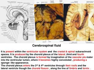
Cerebrospinal Fluid (CSF) Produced at the Choroid plexus of the 4 ventricles by modified Ependymal cells At 90- 150 ml/day is produced CSF flows through the Subarachnoid space Where a volume of 90 – 150 ml is maintained (adults) Reabsorbed at the Arachnoidvillus / granulation to be eventually reabsorbed into the blood
Cerebrospinal Fluid (CSF) Blood Brain Barrier Occurs due to tight fitting endothelial cells that prevent filtration of larger molecules. Controls / restricts / filters blood components Restricts entry of large molecules, cells, etc. Therefore CSF composition is unlike blood’s ** CSF is NOT an ultrafiltrate
Cerebrospinal Fluid (CSF) Blood Brain Barrier Essential to protect the brain Blocks chemicals, harmful substances Antibodies and medications also blocked Tests for those substances normally blocked can indicate level of disruption by diseases: ie meningitis and multiple sclerosis.
Cerebrospinal Fluid (CSF) CSF functions Supplies nutrients to nervous tissues Removes metabolic wastes Protects / cushions against trauma
Cerebrospinal Fluid (CSF) Four major categories of disease Meningeal infections Subarachnoid hemorrhage CNS malignancy Demyelinating disease
Cerebrospinal Fluid (CSF) Indications for analysis To confirm diagnosis of meningitis Evaluate for intracranial hemorrhage Diagnose malignancies, leukemia Investigate central nervous system disorders
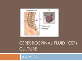
Cerebrospinal Fluid (CSF) Specimen collection and handling Routinely collected via lumbar puncture between 3rd & 4th, or 4th & 5th lumbar vertebrae under sterile conditions Intracranial pressure measurement taken before fluid is withdrawn.
Cerebrospinal Fluid (CSF) Specimen collection and handling Tube 1 – chemistries and serology Tube 2 – microbiology cultures Tube 3 – hematology Testing considered STAT Specimen potentially infectious
Cerebrospinal Fluid (CSF) Specimen collection and handling If immediate processing not possible Tube 1 (chem-sero) frozen Tube 2 (micro) room temp Tube 3 (hemo) refrigerated
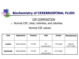
Cerebrospinal Fluid (CSF) Appearance Normal - Crystal clear, colorless Descriptive Terms – hazy, cloudy, turbid, milky, bloody, xanthrochromic Often are quantitated – slight, moderate, marked, or grossly. Unclear specimens may contain increased lipids, proteins, cells or bacteria. Use precautions. Clots indicate traumatic tap Milky – increased lipids Oily – contaminated with x-ray media
Cerebrospinal Fluid (CSF) Appearance Xanthrochromic – Yellowing discoloration of supernatent (may be pinkish, or orange). Most commonly due to presence of ‘old’ blood. Other causes include increased bilirubin, carotene, proteins, melanoma
Cerebrospinal Fluid (CSF) Appearance Clots – indicates increased fibrinogen & usually due to traumatic tap, but may indicate damage to blood-brain barrier. (see below) Pellicle formation in refrigerated specimen associated with tubercular meningitis. Pellicle formation - picture at right (pellicle in L. tube, R is normal) Milky – increased lipids Oily – contaminated with x-ray media
Traumatic collection vs cerebral hemorrhage Cerebral hemorrhage Even distribution of blood in the numbered tubes Clot formation possible Xanthrochromicsupernatent – RBCs must have been in CSF @ 2+ hours - D-dimer, fibrin degradation product from hemorrhage site Microscopic presence of erythrophages, or siderophages, Hemosiderin granules
Cerebrospinal Fluid (CSF) Expected results Normally 0 RBCs/uL regardless of age WBCs Adult – up to 5 mononuclear WBCs/uL Newborn – up to 30 mononuclear WBCs/uL Children (1-4) - up to 20 mononuclear /uL Children (5+) – up to 10 mononuclear / uL Increased numbers = Pleocytosis
Cerebrospinal Fluid (CSF) WBC counts 3% acetic acid can be used to lyse RBC Methylene blue staining will improve visibility
Cerebrospinal Fluid (CSF) Correction of WBC count for traumatic tap contamination. Uses ratio of WBCs to RBCs in blood and compares it to same ratio (WBC/RBC) in CSF If patient’s peripheral cell counts are normal, can subtract 1 WBC for each 700 RBCs counted in CSF. Great chance for considerable error, makes this of little value.


![CEREBRAL CIRCULATION AND CEREBROSPINAL FLUID [CSF]](https://cdn2.slideserve.com/4005143/slide1-dt.jpg)