Download
1 / 6

E N D
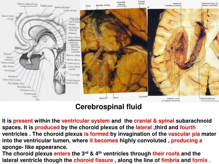
Cerebrospinal fluid It is present within the ventricular system and the cranial & spinal subarachnoid spaces. It is produced by the choroid plexus of the lateral ,third and fourth ventricles . The choroid plexus is formed by invagination of the vascular pia mater into the ventricular lumen, where it becomes highly convoluted , producing a sponge- like appearance. The choroid plexus enters the 3rd & 4th ventricles through their roofs and the lateral ventricle though the choroid fissure , along the line of fimbria and fornix .
Most of the C. S. F. is produced by the choroid plexus of the lateral ventricle. Then it flows through the interventricular foramen ( Foramen of Monro ) into the 3rd ventricle . By way of the cerebral aqueduct to the 4th ventricle. CSF leaves the ventricular system though 3apertures of the 4th ventricle to enter the subarachnoid space. Most passes through the median aperture (of Magendei) to enter the cisterna magna ( It is an enlarged area of the subarachnoid space ) , located between the medulla & cerebellum. Lesser amounts flow through the lateral apertures (of Luschka) to enter the subarachnoid space in the region of the cerebellopontine angle. From these sites , the majority of CSF flows superiorly , round the cerebral hemispheres Small amount passes to central canal of medulla oblongata. Then to central canal of spinal cord.
CSF is reabsorbed into the venous system by passing into the dural venous sinuses , especially the superior sagittal and transverse sinuses. Along the sinuses are located arachnoid villi which consist of invaginations of arachnoid mater through the dural wall and into the lumen of the sinus. Reabsorption occurs at these villi because 1- The hydrostatic pressure (of ionates ) in the subarachnoid space is higher than that in the sinus lumen 2- The greater colloid osmotic pressure (of albumin , proteinate ) of the venous blood compared with CSF.
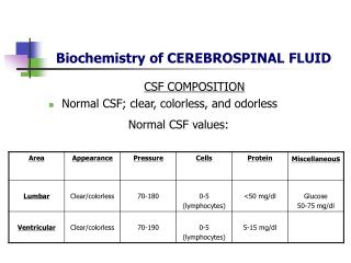
So, CSF is produced by an active secretory process and by passivediffusion . CSF is a colourless and clear fluid containing little protein and few cells, (lymphocytes ) , glucose ; chloride . The volume of CSF in the combined ventricular and subarachnoid spaces is 150 ml . It is in a rate sufficient to fill these spaces several times each day . Functions of CSF 1-Protects the central nervous system from trauma. 2- Provides mechanical support for brain 3- Nourishes the CNS 4- Remove metabolites from the CNS 5- Serves as a pathway for pineal secretions to reach the pituitary gland
With age , the arachnoid villi become hypertrophied to form arachnoid granulations .
Hydrocephalus Obstruction of the flow of CSF within the ventricular system ( by tumors ) Or the subarachnoid space ( by adhesions ) following head injury or meningitis, lead to a rise in the fluid pressure causing swelling of the ventricles (Hydrocephalus ) There is headaches , unsteadiness and mental impairment . Swelling of the optic discs ( Papilloedema ) is seen on ophthalmoscopy. Decompression of the dilated ventricles is achieved by inserting a shunt connecting the ventricles to the jugular vein .


![CEREBRAL CIRCULATION AND CEREBROSPINAL FLUID [CSF]](https://cdn2.slideserve.com/4005143/slide1-dt.jpg)