Download
1 / 42
430 likes | 449 Views
HYPOXIC-ISCHEMIC-ENCEPHALOPHATY. Prof.Maria Stamatin MD,PhD CUZA – VODA Clinical Hospital of Obstetrics & Gynaecology Iasi,NICU. HYPOXIC-ISCHEMIC-ENCEPHALOPHATY. HYPOXIC-ISCHEMIC-ENCEPHALOPHATY (HIE) Brain injury occurs when prolonged hypoxia overwhelms the compensatory mechanisms.

E N D
HYPOXIC-ISCHEMIC-ENCEPHALOPHATY Prof.Maria Stamatin MD,PhD CUZA – VODA Clinical Hospital of Obstetrics & Gynaecology Iasi,NICU
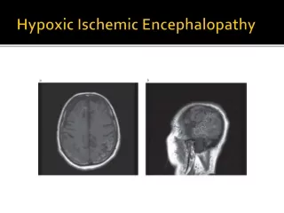
HYPOXIC-ISCHEMIC-ENCEPHALOPHATY HYPOXIC-ISCHEMIC-ENCEPHALOPHATY (HIE) • Brain injury occurs when prolonged hypoxia overwhelms the compensatory mechanisms. • The therm perinatal hypoxic ischemic brain injury includes all varieties of neurological cellular damage. • The incidenceof H.I.E. is about 1-3%, according with frequency of perinatal asphyxia and with possibilities of a succesfull resuscitation. • The link between perinatal asphyxia and brain damage is presented in the next figure:
HYPOXIC-ISCHEMIC-ENCEPHALOPHATY Perinatal Asphyxia HYPERCAPNEEA HYPOXIA ACIDOSIS initially BP REDISTRIBUTION OF CARDIAC OUTPUT LOSS OF CEREBRAL AUTOREGULATION DECREASE BLOOD PRESSURE AND CEREBRAL FLOW INCREASE CEREBRAL BLOOD FLOW HEMORRHAGE BRAIN INJURY
HYPOXIC-ISCHEMIC-ENCEPHALOPHATY • Following the onset of asphyxia, cardiac output is redistributed, so that a large proportion of blood enters in brain, resulting in a 30-175% increase in cerebral blood. When the hypoxic-ischemic insult is prolonged, this homeostatic mechanism fails, cardiac output falls, with systemic hta and reduced cerebral blood flow. • Normal, brain vascularization can compensate for decreased cerebral perfusion by rapid dilatation of the smaller vessels, so that cerebral blood flow is maintained relatively constant as long as blood pressure is kept within normal range. • The constancy of cerebral blood flow in the face of fluctuations in systemic blood pressure is termed autoregulation. Hypoxia, hypercapneea acidosis hypoglycemia may impair cerebral autoregulation.
HYPOXIC-ISCHEMIC-ENCEPHALOPHATY • In the preterm infant, the lower limits of autoregulation are very close to the mean systemic arterial pressure, so that the preterm infant is unable to compensate for relatively small drops in blood pressure. • The exact mechanism of tissue damage in HIE is still unclear. • Under experimental condition, the more severe the brain damages,the more extensive and prolonged the associated edema compressionon cerebral tissue decrease cerebral flow ischemia.
HYPOXIC-ISCHEMIC-ENCEPHALOPHATY • On a molecular level within 15 to 90 seconds after the onset of asphyxia, the neuronal membrane begins to change; if anoxia persists, all cells undergo a rapid and marked depolarization with complete loss of a membrane potential(an influx of Na, CI, and calcium and an eflux of K). • Membrane depolarisation induce the release of the excitotoxic neurotransmitters, which has the ability to mediate hypoxic brain injury (how excitotoxins induces neuronal death is still unclear .
HYPOXIC-ISCHEMIC-ENCEPHALOPHATY • Brain glucose and glycogen also decrease rapidly with asphyxia so that hypoglycemia, by hastening the depletion of energy stores, contributesto asphyxial brain damage. • The severity of brain injury correlates closely with three stages ofclinical features (by Sarnat&Sarnat).
HYPOXIC-ISCHEMIC-ENCEPHALOPHATY • Infants in stage I are irritable, in a hyperalert state, in which they have the eyes open, with a "worried" facial appearance and some degree of feeding difficulties. They seem hungry and respond excessively to stimulation. Tremor, especially when is provoked by abrupt changes of limb position or tactile stimulation, can resemble seizures. • If hypoxic ischemic insult is greater will leads to clinical features of stage II. Infants are lethargic or obnubilated, with delayed or incomplete responses to stimuli. Focal or multifocal seizures are common. • Severely asphyxiated infants develop signs of stage III of HIE.The infant is markedly hypnotic, the sucking and the swallowing reflexes are absent producing difficulties in feeding.
HYPOXIC-ISCHEMIC-ENCEPHALOPHATY (HIE) • A variety of respiratory abnormalities can be encountered. These include a failure to initiate breathing after birth, tachypneea and dispneea in the absence of pulmonary or cardiac disease. • The value of Apgar score in terms of indicating asphyxia is limited but Apgar score is very important in evaluation of latter lesion.
DIAGNOSIS of HIE • DIAGNOSIS: The diagnosis of HIE is based on the following: 1. History of intrauterine distress; 2. History of an abnormal neonatal course; 3. Neurological exam of the newborn 4. Laboratory studies such as: - CSF exams - EEG exams - Ultrasonography of the brain & detected cerebral flow - CT-scan - Enzymatic determination.
HYPOXIC-ISCHEMIC-ENCEPHALOPHATY History of intrauterine distress: Evidence of intrauterine distress includes: • an alteration of fetal heart rate pattern, • the passage of meconium • abnormalities in fetal acid-base status • infants SGA. History of an abnormal neonatal course: • An abnormal neonatal course is the most important diagnosticfeature of perinatal asphyxia, which has been sufficiently severe tocause neurological deficits. • This includes delayed or impaired respiration requiring resuscitative measures such as endotracheal intubation and assisted ventilation.
LABORATORY A.Laboratory findings. • Examination of CSF can also provide evidence for perinatal asphyxia in that the concentration of CSF protein can be elevated (normal values are < 90mg/dlfor term neonate and 115 mg/dl for premature; values above 150 mg./dl are considered abnormal). A normal CSF does not exclude perinatal asphyxia. • EEG-it's value in exploration of preterm and term babies is limited because it didn't exist a basal rhythm. • Ultrasonography of brain has become the major tool for newborn evaluation. and for predicting the outcome of the asphyxiated infant.
LABORATORY • CT scan has wide diagnostic application to babies with neurological disease. In premature baby, CT scan is complicated by the poor visualization of the ventricular system owing to its small volume in relation to brain parenchyma. • Determination of intracranial pressure • Determination of cerebral blood flow (normal values are 40-80ml/100 mg cerebral tissue/min.) • Enzymatic determinations (CK) - creatine kinase increases in first hours after birth, almost in the same time with destruction of haemato-encephalic barrier.
TREATMENT Treatment • The prevention of HIE is largely the task of the obstetrician. • It must be done: 1. the recognition of risk factors, 2. fetal monitorization during labor 3. adequate resuscitation 4. prevention of hypoxemia and hypercapnia • Recognizing during labor of a fetus with hypoxia and acidosis represent and indication for C-section.
TREATMENT Several methods have been suggested to prevent thecomplication of HIEor to reduce its severity: • Administration of phenobarbital sodic( 20 mg/kg-attack dose) in order to treat the convulsions and also appears to reduce the incidence of severe IVH. • Treatment of hypoglycemia • Carefull monitorize: weight,electrolytes values and diuresis .Neither manitol or corticoids are effective in the management of postasphyxial cerebral edema.
PROGNOSIS • Prognosis of HIE • 40-70%-normal evolution • 20-40%-sequelae • 50% - convulsions • 10-20% - mortality
SEQUELS • Among the sequels are: 1. Spastic palsy if both hemispheres are affected + mental retard and convulsions 2. Periventricular leukomalacia (ischaemic necrosis of periventricular white matter) 3. Sensorial and intellectual deficit of various degree. 4. Dystonia and athetosis if basal ganglia are damaged 5. Spastic hemiparesis and ataxia in lesions ofcerebellum
INTRACRANIAL HEMORRHAGE • Intracranial hemorrhage have a become a major source of morbidity and mortality in prematures neonate, especially those weighing less 1500 grams.
INTRACRANIAL HEMORRHAGE • Etiology : • mechanical trauma • concomitent and/or preceding hypoxic ischemiccerebral acute injury • increase in intracranial pressure associated withmajor haemorrage destruction of germinal matrix • focal ischaemia, possible secundary to vasospasm • genetic factors.
INTRACRANIAL HEMORRHAGE • The site of bleeding is determined by the maturity of the infant. • There are four major types of neonatal intracranial hemorrhage: I.Periventricular or intraventricularhemorrhage(PVH/IVH) II. Subdural IlI.Subarahnoid IV.Intracerebellar
INTRACRANIAL HEMORRHAGE I.PVH/IVH • This is the most common form of neonatal intracranial haemorrhage and is specific to prematures, especially to those weighting less than 1500g. • The predisposition of the prematures to PVH/IVHcan be due to presence of a highly vascularized subependimal germinai matrix; further more, the cappilaries of the prematures have less bassement membrane than those of the mature brain. • Abnormalities in the autoregulation of arterioles in premature and distressed term infants impair the childs' response to hypoxia and hypercarbia and thus permit transmission of arterial pressure fluctuation to the fragile periventricular cappillary bed. • Elevation in venous pressure by altering haemodynamics.
INTRACRANIAL HEMORRHAGE Initial breake at capillary jonction level Break of terminal dilated vessels TROMBOSIS Infract+necrosis in white matter Break+ventricular flood Ventricular distension
INTRACRANIAL HEMORRHAGE • Graduating by anatomopathological and clinical criteria: I. grade=subependimal haemorrhage-50% of cases no symptomsor medium hypotonia Il. grade=intraventricular hemorrhage without distension, generallywith a favorable evolution, without sequel III. grade=IVHwith distension and with neuro- logical sequel evolution or death. IV.grade=haemorrhage extented into cerebral parenchyma, with lethal evolution or later major handicaps, such as cerebral palsy.
INTRACRANIAL HEMORRHAGE • Clinical features a) rapid evolution in first 24-48 hfrom birth, when hypoxia take placeduring labor or is secondary to BMH.Neonates presents: coma, arreactive/slow reactive pupilary reaction, neonate is limp, seizures, raised tension of the anterior fontanella, death or hydrocephalus. b) graduate evolution with partial clinical changes, with hypotonia,circular nistagmus; may have good prognosis or present hydrocephalus.
INTRACRANIAL HEMORRHAGE • DIAGNOSIS: Elective method is transfontanelar ultrasonography (indicated to all prematures under 1500g, even without clinical signs) in the first 3 days of life.
TREATMENT • Profilaxy of premature delivery. • Steroids administration to mothers with premature delivery. • Maintain a constant temperature of newborn. • Monitories: respiration,heart rate, PO2, PC02, pH. • Adequate perfusion. • Dynamic ultrasonography. • Phenobarbital administration in first 24 hours after birth,seems todecrease the severity of hemorrhage, due to sedative effect.
TREATMENT • Complicationshydrocephalus. • Lumbar punction in series;extraction of 20 ml of CSF at every punction. • Furosemid administration for decrease CSF formation. • Ventricular stoma with intraventricular catheter (shuntventriculo-peritoneal).
INTRACRANIAL HEMORRHAGE II.Subdural hemorrhage - Is usually the result of mechanical trauma in full-termnewborn,in breech presentation.Typically,the signs begins at birth with coma, respiratory difficulties, and signs of intracranial hypertension.Rarely the onset of neurological deterioration is delayed.Cranial US does not clearly show the subdural hemorrhage,therefore,when is suspected,a CT scan is necessary. - The immediate prognosis for any type of subdural hematomais poor;Outcome following evacuation of hematoma depends on the degree of underlying brain damage.
INTRACRANIAL HEMORRHAGE III.Subrachnoid hemorrhage =break of small vessels from leptomening.It formerly was defined by the presence of over 3000 r.b.c/mmc of CSF. Caused probably by hypoxia with secondary capillary rupture.The N.B. May be asymptomatic or with symptoms like hypertonia,irritability or seizures. IV.Intracerebral hemorrhage-the most severe form and generally with lethal evolution.Usually affects prematures less than 1500g.
NEONATAL SEIZURES • General information A.Seizures are the most frequent major manifestation of neonatal neurologic disorders. B.Reasons why it is important to recognize neonatal seizures, to determinate their etiology and to treat them promptly: • Seizures are usually related to significant illness, sometimes requiring specific treatment(such as meningitis) • Neonatal seizures can last for considerable periods and interfere with important supportive measures (respiration and alimentation) • Seizures in the newborn period may be associated with brain injury.
NEONATAL SEIZURES Pathophysiology • Seizures occur secondary to excesive synchronous electrical discharge- depolarization of neurons within CNS.
NEONATAL SEIZURES The mechanism of seizures is different in neonate versus older children because in neonates myelin deposition, glial proliferation, neuronal migration and axonal-dendritic contacts are incomplete. Types of seizures: • Subtle-eyelid blinking and flutering, sucking, apneea • Generalized tonic-tonic extension of upper extremities/lower extremities-tonic flexion of UE with extension of LE • -term infants clonic jerking • Focal clonic-term infant more frequent than preterm infants; well localized clonic jerking • Myoclonic-either term or preterm;single or multiple jerks of flexion of the upper/lower extremities.
NEONATAL SEIZURES • Jitteriness versus seizures Jitteriness is unique to newborns and the most common causes are: - Hypoxic ischemic encephalophaty; - Hypocalcemia; - Hypoglicemia; - Drug withdrawal; • Clinical featuresJitterinessSeizures 1. Predominant movementtremorclinic jerking 2. Movement cease with+ 0 passive flexion 3. Movement exquisitely + 0 stimulus sensitive 4. Abnormally eyemovement 0 +
NEONATAL SEIZURES Etiology of neonatal seizures(VITAMIN DOC) I.Vascular1 .Hemorrage (1.Periventricular/intraventricular; 2.Subdural; 3.Arterio-venous malformation) 2. Ishemia-asphyxia (1.Trombosis/embolus; 2.Cerebral infraction) Il.Infection (1.Meningitis; 2.Sepsis; 3.TORCH infections) III.Trauma (1. Birth asphyxia; 2. Birth trauma(breech, forceps) IV.Autoimune V.Metabolic(1.Hypoglicemia; 2.HypocCa; 3.HypoMg; 4.Hypo/hyperNa; 5.Pyridoxine dependence) Vl.Inborn errors of metabolism(1. Maple syrup urine disease;2.Methylmalonic acidemia; 3.Nonketotic hyperglycinemia) VII.Neoplasic (Teratomas,gliomas,meduloblastomas) VIII.Drugs (Maternal drugs-narcotics, barbiturics;Teophyline adm.can cause seizures) IX.Other(1. Familial; 2.Idiopathic; 3.HTA; 4.Polycythemia with hyperviscosity ) X.Congenital malformation of CNS
NEONATAL SEIZURES - Diagnosis 1.Hystory-a detailled hystory will help in diagnosing seizure activity. 2.Physical examination-performa complete physical examination, with close attention to the neurological status. 3.Laboratory studies 3.1 Metabolic workup (Serum glucose level;Serum sodium level;Serum ionized and total calcium levels;Serum magnesium level ) 3.2 Infection workup;Hb,Ht and complete blood cell count;Blood, urine and cerebrospinal fluid cultures;Serum immunglobulin M and IgM-specific TORCH titres(the serum titer may be elevated in m TORCH infection) 3.3 Other laboratory studies (Blood gas level to rule out the hypoxia or acidosis;Theophyline level, if the infant is on medication and the toxicity is suspected.) 4.Radiologie and other studies • Ultrasound examination of the head-will confirm periventricular-intraventricular hemorrhage. • CT-scan of the head- can be done to diagnose subarachnoid or subdural hemorrhage.ltmay also reveal a congenital malformation. • Lumbar puncture-the presence of blood in the CSF suggestes IVH • EEG-it is usually not possible to perform EEG during the episode of seizure activity.This study should be done at some time after seizure activity has been documented.ltmay confirme seizure activity and may also be used as a base line study.
NEONATAL SEIZURES - Therapy Therapy - Genaral measures • Once it is determinated that the infant is having seizure, because the other two diagnosis(jitterness and benign myoclonic activity) are benign conditions,immediate management is necessary. • Rule out hypoxia-blood gas measurement-and correct any metabolic acidosis • Check the glucose level- if the paper strip test shows low blood glucose it is acceptable to give 10% glucose, 2-4 ml/kg, by intravenous push, before obtaining results from the laboratory. • Check calcium,sodium and magnesium levels. If these values are low and a metabolic disorder is suspected to be the cause of the seizures, it is acceptable to go ahead and treat the infant before new laboratory values are available. • Anticonvulsivant therapy.If hypoxia and all metabolic abnormalities have been treated or if blood gas and metabolic workup values are normal, start anticonvulsivant therapy. • Give phenobarbital as the first line drug.Initially, 20 mg/kg as loading dose, but up to 40 mg/kg can be given if the seizures have not stopped. • If seizures persist, give phenitoin(Dilantin) at 20 mg/kg/dose. • A trial of pyridoxine with EEG monitoring may be recommended • If seizure still persist, can be used diazepam(continous infusion of 0,3 mg/kg/h).
NEONATAL SEIZURES Specific measures • Hypoxic ischemic injury: caretul observation,prophylactic phenobarbital is used in some institutions, restrict fluids to 60 ml/kg/day. • Hypoglicemia-i.v. perfusion with 10% glucose. • Hypocalcemia-slowly 100-200mg/kg calcium gluconate i.v. • Hypomagnesemia-0,2 meq/kg magnesium sulfate i.v. every 6 hours until magnesium levels are normal and symptoms resolve. • Hyponatremia-calculate sodium defficit and administrate sodium saline perfussion. • Pyridoxine dependence-50-100 mg.i.v. pyridoxine administration.
NEONATAL SEIZURES • Infection-if sepsis is suspected, start administration of a broad spectrum antibiotic until a coplete septic workup and positive results are ready. Duration of therapy-if neonal neurologic exam become normal-discontinue theraphy.If neurologic exam is abnormal, procede an EEG exam, continue phenobarbital with 2-4mg/kg/day as maintenance dose and reevaluate after one month. Prognosis–in relation with neurological disease.If EEG is normal, less than 10% of newborns will have neurological sequelae.If EEG shows moderate abnormalities, a half of newborns will have neurological sequelae and if EEG presents severe abnormalities, more than 90% of babies will have neurological sequelae.