Download
1 / 34
460 likes | 1.2k Views
Neonatology: Hypoxic-Ischemic Encephalopathy, HIE. Main Contents. Clinical definition Etiology/High risk factors Pathogenesis and Pathophysiology Clinical manifestations and diagnostic Neuroimaging Prognosis Clinical Management. Clinical definition.

E N D
Main Contents • Clinical definition • Etiology/High risk factors • Pathogenesis and Pathophysiology • Clinical manifestations and diagnostic Neuroimaging • Prognosis • Clinical Management
Clinical definition Brain damage in Fetus and neonates caused by hypoxic and/or decreasing or abruption of blood flow to brain during perinatal period.
Etiology Almost all the factors causing asphyxia resulting HIE, and • Maternal • Placenta and umbilicus abnormality • Substantial pulmonary, cardiac and CNS disease of the fetus and neonates • Pronged partum • Medication during delivering
High risk factors • Prolonged fetal bradycardia • Repeated late decelerations • Low Apgar scores at 5 minutes or later • Low fetal scalp or cord pH • Requirement for prolonged resuscitation with positive-pressure ventilation
Pathogenesis and Pathophysiology • Change of cerebral blood flow • normal term stable CBF: 50-60ml/min/100g • CBF< 20ml/min /100g, brain damage
Pathogenesis and Pathophysiology • Change of cerebral metabolism • Increase in anaerobic glycolysis • Na +, Ca2 + pump function intracellular ATP exhausted Na +, Ca2 +endosmosis • Irritability amino acid blocking oxidative phosphorylation in mitochondrion • blood stream reperfusion oxygen free radical
Pathogenesis and Pathophysiology • Change of nuropathology • Term baby: cortex infarction gray matter in partes profunda necrosis • Preterm: intraventricular haemorrhage white matter injury • Cerebral inflammation IL-1, TNF- , CKs Cellular apoptosis
Clinical manifestations • Mild • excitation/ irritability • Apparent at 24 hr • No convulsion • normal EEG
Clinical manifestations • Moderate • Convulsion, 50% • with disorder of consciousness • Apparent at 24-48 hr • Deterioration: intensity of anterior fontanelle • coma
Clinical manifestations • Severe • light coma or coma at birth • Irregular respiration and apnea • Convulsion with 12 hr • Poor muscle tone • Intensity of anterior fontanelle • Most die in 1 week • Survivors with severe nerosequelees
HIE的诊断—临床表现 中华医学会儿科学会新生儿学组 2004年11月修订; 长沙 1.胎儿宫内窒息史,严重的胎儿宫内窘迫表现 (胎心<100次,持续5分钟以上;和/或羊水III度污染) 2.出生时有重度窒息:(Apgar评分1分钟≤ 3分) 至5分钟时仍≤5分;或出生时脐动脉血气pH ≤ 7.00; 3、出生后24 小时内出现神经系统表现; 4、排除低钙血症、低糖血症、感染、产伤和颅内出血等引 起的抽搐,以及遗传代谢性疾病和其他先天性疾病所引 起的神经系统疾患。 • 同时具备以上4条者可确诊,第4条暂时不能确定者作为 拟诊病例。
HIE的诊断—脑电图 • 在生后1周内检查 • 脑电图异常程度与临床分度基本一致 • 脑电图异常表现: 脑电活动延迟 (落后于实际胎龄), 背景活动异常 (以低电压和爆发抑制为主) • 振幅整合脑电图 (aEEG) 中华医学会儿科学会新生儿学组 2004年11月 长沙修订
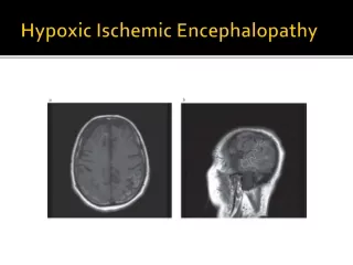
HIE的诊断—影象学检查 中华医学会儿科学会新生儿学组 2004年11月修订; 长沙 • 头颅B超 可在HIE病程早期 (72小时内) 开始检查 有利于了解脑水肿、基底神经节丘脑损伤 和脑动脉梗死等病理改变 • CT 生后4-7天为宜 • MRI 对HIE病变性质与程度评价方面优于CT
Neuroimaging Cerebral edema US
Neuroimaging Cerebral edema CT MRI
Neuroimaging • injury in Hypothalamus and Basal ganglia US
Neuroimaging injury in Hypothalamus and Basal ganglia MRI CT
Neuroimaging injury in Area adjacent to the sagittal CT MRI
Neuroimaging US 早期回声增强 Cerebral artery Infarction in terms
Neuroimaging Cerebral artery Infarction in terms CT MR I
Neuroimaging PVL in premature US
Neuroimaging PVL in premature CT MRI
Neuroimaging Punctate encephalon haemorrhage MRI
Severity and diagnosis 中华医学会儿科学会新生儿学组 2004年11月修订; 长沙 • Mild • Irritability, normal tone.. • Moro’s: ; Sucking: normal • normal respiration,no convulsion • Moderate • Oppressed,muscle tone ,Moro’s and Sucking • convulsion。>7-10d, may have sequelae • severe • coma,frequently convulsion • irregular respiration or apnea. respiration failure. very high death rate • Survivors usually have sequelae
Prognosis • Mild and Moderate Recovered <5d, good outcome • Middle >7d,or Severe worse outcome
Clinical Management • For an asphyxiated newborn: • immediate maintenance of ventilation and perfusion • control of seizures • maintenance of metabolic homeostasis, especially blood glucose levels to avoid additional cerebral insult
Clinical Management • Maintenance of adequate ventilation: • Avoidance of hypoxemia and hypercapnia • To avoid systemic hypotension • cerebral perfusion • Prevention of fluid overload: • current data in human newborns do not provide convincing evidence that supports the use of antiedema therapy • Maintenance of normoglycemia
Clinical Management • Control seizures • begin with a loading dose of phenobarbital (20mg/kg) ,IV • followed by additional 5-mg/kg, total dose 40 mg/kg • For refractory seizures: • lorazepam by IV may be indicated • Recent recommendations emphasis: • brief duration of treatment; possible deleterious effects of anticonvulsants on the developing nervous system.
Clinical Management • Cool Cap (Selective Head Hypothermia Therapy) • Multi-center trial: • US, Canada, UK and New Zealand: 25 • Sample: trial/control=116/118 • Apgar<=6/5min+Cord arterial ph <7.1 • clinical HIE+EEG abnormal • aEEG severe: (n=46):not effective • aEEG Moderate : (n=172); showed protective Gluckman PD, Cool Cap trial group. Lancet 2005
Clinical Management • Cool Cap (Selective Head Hypothermia Therapy) • aEEG Moderate : (n=172); showed protective • Death rate: • severe neromotion disabled 48% vs 66% p=0.02 • Bayley MDI: 85 vs 77 p=0.04 • Bayley PDI: 90 vs 85 p=0.047 Gluckman PD, Cool Cap trial group. Lancet 2005
Clinical Management • Whole body Hypothermia • NIH Neonatal Network,US • Multi-center:16, sample:208 • Results; • Death: 24%(H) vs 36% p=0.08 • middle or severe disabled • 45%(H) vs 62%(N) p=0.01 (OR: 0.72, 95% CI 0.55-0.93) Shankaran et al:National Institute of Child Health and Human Development Neonatal Research Network. Whole -body hypothermia for neonates with hypoxic-ischemicencephalopathy.NEJM 2005 Oct 13;353(15):1574-84.
Summery • HIE is the major cause of the neonatal death • Asphyxia and ischemia hypoxemia in perinatal resulting in HIE • Diagnosis based on clinical manifestation and may combined with Neuroimaging • Though there are some therapies for HIE treatments for HIE is still not as effective as expected