Download
1 / 47
470 likes | 745 Views
POOR CONFORMATION: HIP DYSPLASIA. YOUNG DOGS 5-8 mos AND MATURE ANIMALS WITH CHRONIC DISEASE. POOR CONFORMATION: HIP DYSPLASIA. POOR CONFORMATION: HIP DYSPLASIA. Acetabular vs. Femoral hip dysplasia. Poor conformation combined with genetic, environmental and nutritional factors.

E N D
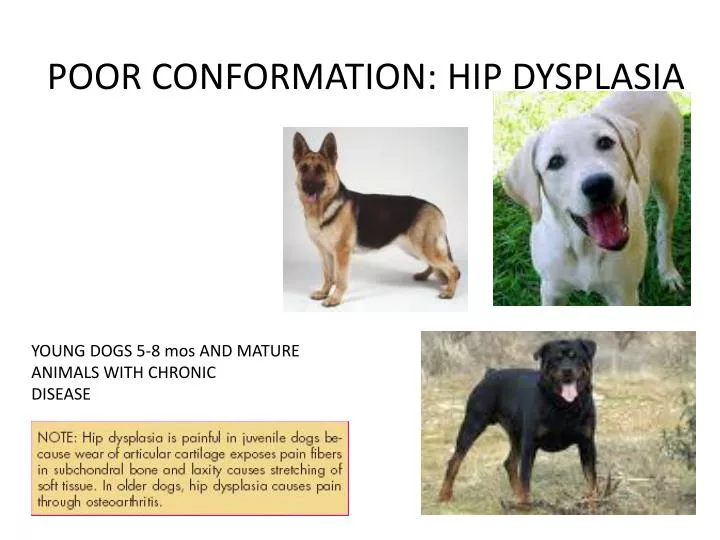
POOR CONFORMATION: HIP DYSPLASIA YOUNG DOGS 5-8 mos AND MATURE ANIMALS WITH CHRONIC DISEASE
POOR CONFORMATION: HIP DYSPLASIA Acetabular vs. Femoral hip dysplasia Poor conformation combined with genetic, environmental and nutritional factors
POOR CONFORMATION: HIP DYSPLASIA • PHYSICAL EXAM FINDINGS • Pain on palpation of hips • Joint laxity (positive ortolani sign) – early disease – subluxation of hip • Crepitus • Decreased ROM of hip joints • Atrophy of thigh muscles • Hypertrophy of shoulder muscles
POOR CONFORMATION: HIP DYSPLASIA http://www.youtube.com/watch?v=2rRKDheDrLs&NR=1 http://www.youtube.com/watch?v=SHCIT87jY0M&feature=related
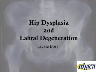
Hip Dysplasia: Radiographic view ventrodorsal view of the pelvis with rear limbs extended symmetrically and rotated inward to center the patellae over the trochlear grooves For standard Orthopedic Foundation for Animals–type radiographs to evaluate hip conformation, extend the hips and internally rotate the tibias until the patella lies directly over the trochlear grooves. Be sure the pelvis is straight, with symmetric obturator foramina.
POOR CONFORMATION: HIP DYSPLASIA and OFA CERTIFICATION "normal" radiographically may be further classified as excellent, good, fair, or near normal
HIP DYSPLASIA and OFA CERTIFICATION dysplasia are categorized as mild, moderate, or severe
HIP DYSPLASIA TREATMENT NSAIDs NEUTRICEUTICALS/CHONDROPROTECTIVE AGENTS
HIP DYSPLASIA TREATMENT: MEDICAL MANAGEMENT • Aspirin or buffered aspirin: 10-25 mg/kg q 8-12 hr or as needed: Discontinue if vomiting occurs. • Carprofen (Rimadyl): 2 mg/kg PO q 24 hr Deracoxib (Deramaxx): For chronic dosing use 1-2 mg/kg PO q 24 hr as needed. • Etodolac (EtoGesic): 10-15 mg/kg PO q 24 hr • Firocoxib (Previcox): 5 mg/kg PO q 24 hr. Do not use in puppies less than 7 months of age or in dogs weighing less than 7 pounds. • Meloxicam: 0.2 mg/kg first dose; then 0.1 mg/kg thereafter q 24 hr PO. • Tepoxalin (Zubrin): 20 mg/kg PO q 24 hr x 1 treatment; then 10 mg/kg PO q 24 h. This is similar to carprofen and ketoprofen.
HIP DYSPLASIA TREATMENT: MEDICAL MANAGEMENT • Polysulfatedglycosaminoglycan (Adequan): 5 mg/kg IM every 3-5 days for 8 injections, followed by an injection every 1-2 months for maintenance: • Polysulfatedglycosaminoglycans prevent cartilage breakdown by inhibiting the enzymes of cartilage degradation during inflammation. • Discontinue use if there is no improvement after the third week. Caution, may increase bleeding time. • Cosequin: 1-2 regular strength capsules PO q 24 hr for smaller dogs and 2-4 capsules of double strength for larger dogs: • Note that dose is based primarily on empiricism and manufacturer's recommendations. • Adverse effects have not been reported although hypersensitivity is possible. • Cosequin is a brand name for glucosamine HCL combined with chondroitin sulfate which may stimulate synthesis of synovial fluid, inhibit degradation, and improve healing of articular cartilage.
Hip Dysplasia – Treatment • Surgical • Total hip replacement • Salvage procedure in mature dogs with severed DJD unresponsive to medical Tx • Pain free in 90% of cases • Unilateral replacement provides acceptable function in 80% of cases • Excision Arthroplasty or Femoral Head Ostectomy • Forms “false” joint • Removal of femoral head and neck to prevent joint pain • Salvage procedure when medical treatment not working and other sx too expensive • Best - < 20#; good musculature • Abnormal gait • Triple Pelvic Osteotomy
Arthroscopy – Juvenille patients A, Ventrodorsal radiograph of an immature dog with subluxation of the femoral heads and minimal evidence of DJD, typifying a candidate for triple pelvic osteotomy. B, Ventrodorsal radiograph of a dog with advanced hip dysplasia and osteophyte formation. This dog may be a candidate for total hip replacement or femoral head ostectomy if clinical signs cannot be managed medically. Arthroscopic view of a normal hip joint
HIP DYSPLASIA TREATMENT TRIPLE PELVIC OSTEOTOMY
HIP DYSPLASIA TREATMENT FEMORAL HEAD OSTECTOMY “False joint” forms from scar/fibrous tissue
HIP DYSPLASIA TREATMENT artreality.com www.kahalapethospital.com/yahoo_site_admin/as...
JUVENILE PUBIC SYMPHYSIODESIS • Juvenile pubic symphysiodesis (JPS) surgery is a prophylactic procedure performed in puppies 10 to 20 weeks of age that have been diagnosed with hip dysplasia • causes premature closure of the cranial pubic symphysis PennHip distraction view of a Labrador puppy at 14 weeks. The DI is 0.55. The same dog at 50 weeks (36 weeks post-JPS).
Hip Dysplasia – Client Info • Weight control important to decrease load on painful joint • Swimming excellent activity • Lameness may occur due to surgical shortening of the affected limb • Physiotherapy – decreases joint stiffness, helps maintain muscle integrity • Joint degeneration progressive • May be heritable – do not breed • Special diets designed for fast growing dogs may decrease severity
LEGG-CALVE-PERTHES DISEASE YOUNG, SMALL BREEED DOGS http://www.youtube.com/watch?v=_vvtprqhyoI
LEGG-CALVE-PERTHES DISEASE May also be considered a Developmental disorder
LEGG-CALVE-PERTHES DISEASE: TREATMENT FEMORAL HEAD AND NECK EXCISION FHNE
OSTEOCHONDRITIS DISSECANS FAILURE OF THE LOWER LAYERS OF ARTICULAR CARTILAGE TO MATURE INTO BONE RESULTS IN THICKENED CARTILAGE THAT IS PRONE TO INJURY
OSTEOCHONDRITIS DISSECANS CARTILAGE FLAP OF THE SCAPULOHUMERAL JOINT WHICH IS THE MOST COMMON LOCATION
OSTEOCHONDRITIS DISSECANS CARTILAGE FLAP IN THE STIFLE JOINT
OSTEOCHONDRITIS DISSECANS http://www.youtube.com/watch?v=_bJqjqh5a2A
INFLAMMATORY CONDITIONS: PANOSTEITIS LARGE, MEDIUM BREEDS AT A YOUNG AGE 6-18 MONTHS CAUSE: UNKNOWN, BUT THERE ARE MANY SUSPECTED CONTRIBUTORS
PANOSTEITIS • PELVIS • LESIONS OF PANOSTEITIS • FEMUR INFLAMMATION IN THE MARROW CAVITIES OF LONG BONES THAT LEADS TO PAIN, LAMENESS, AND FEVER
PANOSTEITIS INCREASED MEDULLARY OPACITY
PANOSTEITIS TREATMENT OF PANOSTEITIS INCLUDES NSAIDS AND CAGE REST THIS DISEASE IS SELF-LIMITING AND HAS A GOOD PROGNOSIS!
LUXATIONS HX OF TRAUMA, ACUTE LAMENESS, NON WEIGHT BEARING, POSSIBLE SWELLING OVER THE HIP
LUXATIONS CRANIODORSAL LUXATION IS THE MOST COMMON TYPE
LUXATIONS THE EHMER SLING IS USED AFTER CLOSED REDUCTION OF THE LUXATED HIP JOINT; THE DOG SHOULD BE CONFINED FOR 7-10 DAYS
TUMORS OF THE BONE MOST COMMON IN LARGE BREED MALE DOGS OLDER THAN 7 YRS OF AGE THE DISTAL RADIUS IS THE MOST COMMON LOCATION
TUMORS OF THE BONE 85%-90% OF BONE CANCER IN DOGS IS OSTEOSARCOMA
TUMORS OF THE BONE OSTEOSARCOMA TENDS TO OCCUR AWAY FROM THE ELBOW AND TOWARDS THE KNEE
TUMORS OF THE BONE METASTASIS OF OSTEOSARCOMA TO THE LUNGS; THERE IS USUALLY ALREADY SOME MICROSCOPIC SREAD OF TUMOR BY THE TIME IT IS DIAGNOSED
TUMORS OF THE BONE AMPUTATION OF THE AFFECTED LIMB ALONG WITH CHEMOTHERAPY IS A COMMON TREATMENT PLAN SURVIVAL TIME IS ~12 MONTHS EVEN WITH AGGRESSIVE THERAPY
MYOPATHIES: MASTICATORY MUSCLE MYOSITIS (aka atrophic myositis, eosinophilic myositis) CLINICAL SIGNS INCLUDE ELEVATED 3RD EYELIDS, ATROPHY OF THE JAW MUSCLES, AND INABILITY TO OPEN THE MOUTH (TRISMUS)
MYOPATHIES: FELINE POLYMYOPATHY CERVICAL VENTROFLEXION OF THE NECK 2° TO HYPOKALEMIA