Download
1 / 42
700 likes | 1.57k Views
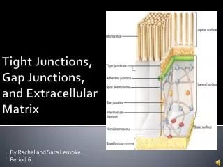
Chapter 19: Cell junctions and the extracellular matrix. Know the terminology: Cadherins, integrins, tight junction, desmosome, adhesion belt, hemidesmosome, gap junction, connexin/connexan,

E N D
Chapter 19: Cell junctions and the extracellular matrix Know the terminology: Cadherins, integrins, tight junction, desmosome, adhesion belt, hemidesmosome, gap junction, connexin/connexan, Basal lamina, connective tissue, fibroblast, proteoglycan, GAG (glycosaminoglycan), hyaluronin, aggrecan, collagen, fibronectin,
Overview Multicellular organisms combine cells into tissues. Extracellular matrix is a complex network of macromolecules (proteins, glycosaminoglycans, proteoglycans) secreted by cells Specialized ECMs include connective tissue, basal lamina, exoskeletons, cartilage/bones, Tissues are formed from cell-cell connections and cell-matrix connections.
Outline How are cells organized into tissues? I. Cell junctions -types of cell junctions -tight junctions, adherans junctions, desmosomes, focal adhesions, hemidesmosomes, gap junctions II. Extracellular matrix proteins -types of ECM macromolecules -synthesis and properties of hyaluronan, aggrecan, collagen, fibronectin
Cell junctions Three types of cell junctions: 1. Occluding junctions: seal cells together into sheets (forming an impermeable barrier) 2. Anchoring junctions: attach cells (and their cytoskeleton) to other cells or extracellular matrix (providing mechanical support) 3. Communicating junctions: allow exchange of chemical/electrical information between cells
Occluding junctions Example: Tight junctions of intestinal epithelium
Tight junction Each cell possesses integral membrane proteins that bind to similar proteins in the adjacent, forming a continuous “weld”
Anchoring junctions Integral membrane proteins connect a cell’s cytoskeleton to another cell or extracellular matrix
Anchoring junctions Integral membrane proteins connect a cell’s cytoskeleton to another cell or extracellular matrix
Anchoring junctions Cytoskeletal fibers (MF, intermediate filaments) connect to a Membrane protein receptor which attaches to another protein in either: -the extracellular matrix or -another cell membrane
Cadherins and desmosomes Cell to cell connections are mediated by cadherins. These receptors extend out from the cell, binding to other cadherens
Cadherins participiate in adherens junctions Under the cell membrane, contractile fibers of microfilaments connect to cell membrane proteins called cadherins They surround the cell, forming a belt
Desmosomes Cadherins attach to intermediate filaments via anchoring proteins: a desmosome Cadherins can also form localized spot connections
Cells-to-ECM attachments: Focal adhesions and hemidesmosomes Cytoskeletal fibers attach to transmembrane receptors (integrins) that are attached to extracellular matrix components Focal adhesions use MF Hemidesmosomes use IF
Gap junctions Gap junctions allow cells to exchange electrical and/or chemical signals Composed of proteins that form channels that allow small molecules to pass. Subunits of these channels are connexins that are assembled together to make connexons. The connexons from 2 cells join together to make a gap junction.
Regulation of connectivity When might a cell want to alter its connections to other cells? How do cells alter these connections? -alter the profile of cytoskeletal connections, receptors, and extracellular matrix -alter the binding affinity of receptors -many are Ca2+ dependent -many are affected by protein kinases
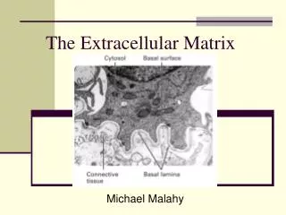
Extracellular matrix A network of proteins, glycosoaminoglycans (GAGs) and combinations of the two (proteoglycan) found in the extracellular space All ECM components are secreted by cells Most cells can secrete elements of the ECM but many ECMs are built by fibroblasts Examples of ECM: -connective tissue, basal lamina, cartilage, bone, plant/fungi cell walls, myelin sheath,
Components of the extracellular matrix Proteins/glycoproteins (secreted by co-translational import) differ in physical properties (e.g. size, flexibility) and are able to bind to different combinations of macromolecules on cell membranes (e.g. integrins, cadherins) and other ECM elements Examples include: -collagen, elastin, fibronectin, laminin
Components of the extracellular matrix Proteins can provide elastic properties in many tissues (e.g. elastin)
Components of the extracellular matrix GAGs differ in physical properties (e.g. size, flexibility, hydration). Composed of repeating sugar + amino sugar units (e.g. N-acetylglucosamine, N-acetylgalactosamine) They occur in long strings, often attached to proteins Examples include: -hyaluronan, chondroitin sulfate, heparan sulfate, keratan sulfate
GAGs GAGs attract a great deal of water. Hydroxyl groups form hydrogen bonds, and the many negative charges attract clouds of cations (Na+) that induce an osmotic movement of water. These hydrated gels resist compression (useful for joints).
Components of the extracellular matrix Proteoglycans (made of both proteins and GAGs) also differ in physical properties Synthesized in Golgi prior to secretion In addition to structural roles, proteoglycans can also bind hormones (e.g., inflammatory chemokines, FGF, TGFb) to alter cell signaling pathways Examples include: -decoran, aggrecan (the main component of cartilage)
ECM proteins, GAGs, and proteoglycans Green-protein Red-GAG
Hyaluronan Very simple GAG, consisting of 10,000+ repeats of glucuronic acid and N-acetylglucosamine Spun directly from cell membranes by a surface enzyme complex Attracts water and fills spaces between cells with non-compressible gel (found around joints) Some cells secrete it to isolate themselves from other cells (e.g. myoblasts). These cells can secrete hyaluronidase to break it down, allowing contact
Hyaluronan Very long macromolecule that hydrates and fills enormous volumes
Aggrecan One of the largest macromolecules, consisting of a core protein with GAGs attached to form a feather-like appearance.
Aggrecan An aggrecan core protein is very large but also binds many (different) GAGs (shown in red)
Aggrecan aggregates Each aggrecan can be attached to a hyaluronan backbone to form an aggrecan aggregate
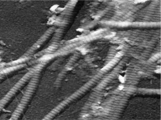
Collagen All multicellular animals possess collagens, often with many different collagen genes Synthesized as pro-collagen monomers (pro-a collagen) Peptide modified by hydroxylation and glycosylation Prior to secretion they self- assemble into trimers Upon secretion the trimers are processed by proteolytic enzymes then assemble into fibrils
Collagen fibers Collagen proteins (trimers) are then cross-linked to form collagen fibers (stiff, not very elastic)
Fibronectin Animals have a single fibronectin gene that can be alternatively spiced into 40+ forms. Different exons are able to bind different proteins/GAGs (e.g. integrins, collagen, etc) Fibronectin dimerizes using 2 similar (not identical) monomers
Control of ECM Cells control the synthesis of ECM by altering gene expression and co-translational import and secretion They also control the degradation of the ECM by secreting and activating/inactivating extracellular enzymes. Most important are a group of serine proteases called matrix metalloproteinases
MMPs Cells that need to migrate must first break down the connections to the ECM (e.g. tissue repair, metastasis of tumors) MMPs can be: -secreted in active form (collagenase) -secreted as inactive form (e.g. plasminogen is inactive until it is modified by “plasminogen activators) -activated when cells stop secreting TIMPs (tissue inhibitors of metalloproteinases)