Download
1 / 103
1.03k likes | 1.56k Views
Chapter 28 Muscular Movement and Support. Striated muscle. Movement is a displacement from one point to another, and occurs at all levels of organisation: atomic, molecular, cellular, organ, organism and population. Locomotion is the movement of a whole organism from one place to another.

E N D
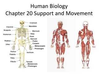
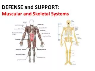
Chapter 28Muscular Movement and Support Striated muscle
Movementis a displacement from one point to another, and occurs at all levels of organisation: atomic, molecular, cellular, organ, organism and population. • Locomotion is the movement of a whole organism from one place to another. • While both plants and animals exhibit movement, only animals carry out locomotion.
There are a number of reasons why animals move from place to place: 1. To obtain food 2. To escape from predators 3. To find a mate 4. To distribute offspring 5. To reduce competition 6. To avoid danger 7. To maintain position 8. To avoid waste products
Small organisms would be advantageous in movement because less energy is needed during locomotion and for support on land. • Plants have to be large in order to have a large surface area for efficient absorption of light, carbon dioxide and water. • Animals must move in order to obtain food, except sessile animals which obtain food by filter feeding in the medium (water) they live. • An aquatic mode of life in these sessile animals also facilitates external fertilization to take place.
Pseudopodia - temporary projections of a cell in amoeba, WBCs. • Movement is achieved in three ways: use of pseudopodia, cilia or muscle. pseudopodium
The plasma membrane is slightly adhesive to its substrate. • Cytoplasm is divided into 2 regions: outer ectoplasm consists of the more rigid plasmagel; inner endoplasm consists of the more fluid plasmasol. pseudopodium
During locomotion, the central core of endoplasm moves forwards, pushing out the tip of the pseudopodium to form the more viscous ectoplasm and flows backwards along the outer portion of the cell. • At the anterior end: gelation –forming jelly, • At the posterior end: solation –forming solution. pseudopodium
Cilia and Flagella • Flagella & cilia are fundamentally similar, the additional length of a flagellum being its main distinguishing feature. • A flagellum produces a wave-like motion usually in one plane, sometimes be spiral. • Commonly the flagellum is at the rear of a cell to propel it forwards. Flagellum
In cilia, the effective stroke is rigid & extends rapidly in a complete arc to present the greatest resistance to propel the cell forwards. • The recovery stroke is more flexible and returns slowly in a folded position with far less resistance against the medium. • The structures of cilia and flagella are remarkably similar in all organisms with the 9 + 2 arrangement of filaments. Cilium
Cilia do not function independently as do flagella, but beat in co-ordinated waves(metachronal rhythm). • The wave of effective strokes begins at some point on the membrane and spreads along its length in a single direction. Cilium
Functions of flagella: 1. Locomotion by flagellates, e.g. Euglena 2. Feeding in sponges 3. Reproduction, e.g. sperms in mammals Functions of cilia: 1. Locomotion by ciliates, e.g. paramecium 2. Feeding in mussels by filter feeding 3. Reproduction, e.g. movement of ova down oviduct 4. Gaseous exchange, e.g. cilia in respiratory tract 5. Transport of nutrients, e.g. central canal & ventricles of CNS
Detailed structure of muscle Diagram to show molecular Interpretation of the EM image Smooth muscle fibre A myofibril nucleus A band M line Z line I band A band I band Z line M line motochondrion
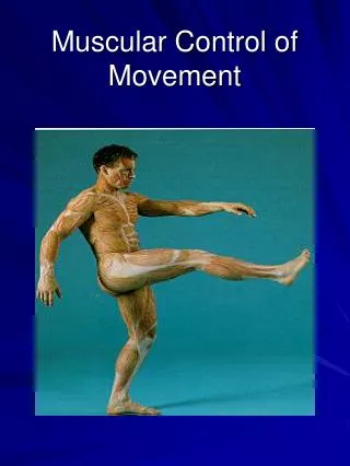
Muscular Movement - muscles are attached to bones to form lever systems for movement - muscle contraction creates a driving force when obtains energy from respiration; when a muscle relaxes, energy is not necessary There are 3 types of muscle: • Skeletal muscle, • Smooth muscles, • Cardiac muscle.
There are 3 types of muscle: Skeletal muscle - muscles attached to bone surfaces - under conscious control (voluntary muscle) - each muscle fibre is multinucleate with striations - produces powerful contractions but becomes fatigued if remains contracted for a long time because lactic acid accumulates in the muscle cells
There are 3 types of muscle: Smooth muscle - found in internal organs - not under conscious control (involuntary muscle) - possesses a central nucleus & is non-striated - produces less powerful contractions but can remain contracted for a long time Cardiac muscle (heart muscle) - found only in heart - not under conscious control (involuntary muscle) - contracts automatically & is regulated by nerves - each muscle fibre is striated, with bridges joining neighbouring muscle fibres - contracts powerfully and does not become fatigued unless the heart dies
28.1 Structure of Skeletal Muscle - an individual muscle is made up of hundreds of muscle fibres; - a muscle fibre is multinucleate with cross striations & is composed of numerousfibrils arranged parallel to one another
- sarcolemma: membrane of muscle fibre sarcomere: each repeating unit of cross striations sarcoplasm: cytoplasm with a system of membranes called sarcoplasmic reticulum
anisotropic band: dark band and isotropic band: light band Z line: central line of each light band; 2 Z lines marks a sarcomere H zone: lighter region in dark band with a central dark line - M line Myosin: thick filaments Actin: thin filaments
28.1.1 The Neuromuscular Junction or end plate - the point where the effector nerve meets a skeletal muscle - many end plates spread throughout a muscle to stimulate a group of fibres called effector (motor) unit to create a rapid and powerful muscle contraction
Striated muscle fibre Motor end plate End plate (400x app.)
Motor neurone Skeletal muscle fibre False colour scanning EM of a neuromuscular junction (3600x app.)
When a nerve impulse is received at the end plate, synaptic vesicles fuse with the end plate membrane and release their acetylcholine. • The transmitter travels across the sarcolemma where it alters its permeability to sodium ions which now rapidly enter, depolarizing the membrane.
Provided the threshold value is exceeded, an action potential is fired in the muscle fibre and the effector (motor) unit served the end plate contracts. • Acetylcholine then breaks down by enzyme to ensure that the muscle is not overstimulated and the sarcolemma becomes repolarized.
28.2 Muscular Contraction - Each muscle is attached to the skeleton at both ends by tendons - Tendons are made of a tough connective tissue (collagen) - Insertion: the end of a muscle (i.e. tendon) attached to a movable bone during muscle contraction Origin: the end of a muscle attached to a fixed bone - Neuromuscular junction: the point where the nerve joins the muscle fibre - When a muscle contracts, it becomes fatter & shorter to pull When a muscle relaxes, it becomes thinner & longer, BUT IT CANNOT PUSH
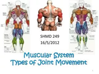
MOVEMENT OF THE FOREARM • Muscles work in pairs for movement of a limb; • They are called antagonistic muscles because they contract and pull in opposite directions; one contracts while the other relaxes
flexor: muscle that contracts and bends the limb • extensor: muscle that contracts and straightens the limb
28.2.1 Mechanism of Muscular Contraction - the sliding filament theory -filaments of actin & myosin slide past one another -actin filaments, hence the Z lines to which they are attached, are pulled towards each other, sliding as they do over the myosin filaments. No shortening of either type of filament occurs.
Appearance of a muscle fibre: 1. The isotropic band becomes shorter 2. The anisotropic band does not change in length 3. The Z lines become close together, i.e. the sarcomere shortens 4. The H zone shortens
Relationship of tropomyosin & troponin to the actin filament
28.2.2 Summary of Muscle Contraction: 1.Impulse reaches the neuromuscular junction 2.Synaptic vesicles fuse with end-plate membrane & release a transmitter (e.g. acetylcholine) 3. Acetylcholine depolarizes the sarcolemma 4. Acetylcholine is hydrolysed by acetylcholinesterase 5. Provided the threshold value is exceeded, an action potential is created in the muscle fibre 6. Ca2+ are released from the T-system & sarcoplasmic reticulum 7. Ca2+ bind to troponin, changing its shape
8.Troponin displaces tropomyosin which has been blocking the actin filament 9.Myosin heads now become attached to the actin filament 10. The myosin head changes position, causing the actin filaments to slide past the stationary myosin ones 11.An ATP molecule becomes fixed to the myosin head, causing it to become detached from the actin 12.Hydrolysis of ATP provides energy for the myosin had to be ‘cocked’ 13.The myosin head becomes reattached further along the actin filament
14. The muscle contracts by means of this ratchet mechanism 15. The following changes occur: (a) I band shortens; (b) Z lines move closer together (sarcomere shortens); (c) H zone shortens. 16. Ca2+ are actively absorbed back into the T-system 17.Troponin reverts to its original shape, allowing tropomyosin to again block the actin filament 18. Phosphocreatine is used to regenerate ATP
28.3 Skeletons and Support Both aquatic and terrestrial organisms need a skeleton to support them against the pull of gravity. Skeleton fulfil 3 main functions: support, locomotion and protection.
28.3.1 Types of Skeleton Hydroskeleton
28.3.1 Types of Skeleton Hydroskeleton - typical of soft-bodied organisms (e.g. earthworm) with liquid secreted and trapped within body cavities - antagonistic muscles are arranged segmentally: circular muscles make the body thinnerbut longerlongitudinal muscles make the bodyfatter but shorter - chaetae on each segment anchor body to substratum to facilitate alternate contractions of both muscles to drive the body forwards with waves of contraction pass along the body
Exoskeleton - an external covering protecting internal organs & providing attachment for muscles in arthropods - three layers: epicuticle (waxy), exocuticle (rigid layer of chitin) & endocuticle (flexible layer chitin) - joints present with flexible parts separating inflexible parts of skeleton - may be impregnated with salts (CaCO3) to give addition strength (crustaceans) - ecdysis overcomes the limitation of growth in these animals