Download
1 / 61
751 likes | 1.67k Views
Normal Radiographic Anatomy of the Thorax. James Grierson BVetMed CertVR CertSAS MRCVS. Normal Radiographic Anatomy of the Thorax. Obtaining a good thoracic radiograph Review Anatomy Case Examples. Obtaining a good thoracic radiograph.

E N D
Normal Radiographic Anatomy of the Thorax James Grierson BVetMed CertVR CertSAS MRCVS
Normal Radiographic Anatomy of the Thorax • Obtaining a good thoracic radiograph • Review Anatomy • Case Examples
Obtaining a good thoracic radiograph • Good quality image improves recognition of normal anatomy • Improves accuracy of diagnosis • Record exposure settings • Allows follow-up to assess disease
Obtaining a good thoracic radiograph • Projections • Lateral and DV minimum • Positioning • Lateral – pull legs forward, place wedge under chest to reduce rotation. • Centre middle of thoracic cavity at caudal point of scapulae, 1/3rd up from sternum • DV – ensure symmetry. Centre midline, just caudal to scapulae
Obtaining a good thoracic radiograph • Collimation • Include entire thoracic cavity. Thoracic inlet cranially and 11th rib caudally. • Dorsally thoracic spine, ventrally sternum • Restraint • General anaesthesia ideal – allows inflation • Sedation • Sandbags and foam wedges • Avoid manual restraint
Obtaining a good thoracic radiograph • Inspiratory films improve image quality • Inflated views if under anaesthesia • Ensure good safety precautions • Inspiratory versus Expiratory
Inspiratory Expiratory
Obtaining a good thoracic radiograph • Film/screen combination • Fastest available – avoids movement blur • Grid • Always for medium/large dogs, not essential in cats • Increases exposure factors • Don’t use if time needs to be increased
Obtaining a good thoracic radiograph • Exposure • High kV to produce low contrast image • Reduces relative opacity of ribs • Low exposure time <0.15secs • High mA (to reduce time) • Toast!!
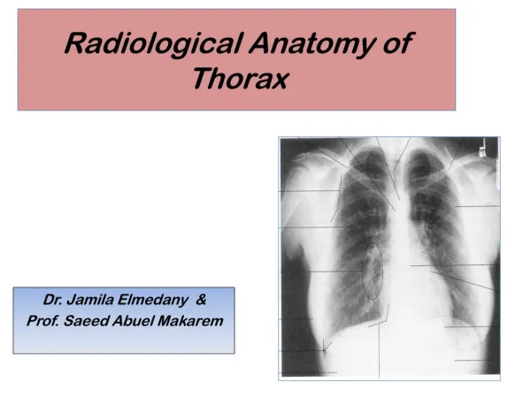
Anatomy • Soft tissues of the thoracic wall • Skin and subcutaneous tissues • Boundaries of the thoracic cavity • Spine, sternum, ribs, diaphragm, thoracic inlet • Pleural space • Mediastinum • Oesophagus, lymph nodes
Anatomy • Airways • Trachea, main stem bronchi • Lungs • Bronchi, bronchioles, alveoli, vessels • Heart and great vessels • Aorta, vena cava, pulmonary artery/vein
Soft tissues of the thoracic wall • Skin and subcutaneous tissues
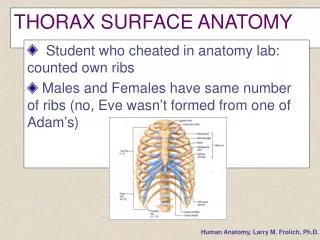
Boundaries of the thoracic cavity • Spine • Sternum • Ribs • Diaphragm • Thoracic Inlet
Manubrium Xiphisternum
Left Crus Right Lateral Stomach Right Crus CVC Heart
Diaphragm – Lateral views LEFT RIGHT L CRUS L CRUS CVC R CRUS HEART R CRUS
Costo-phrenic angle
Pleural space • Not radiographically visible • Seen if contains fluid or air
Mediastinum • Non-event radiographically • Oesophagus • Not normally visible • Seen if gas filled – tracheal stripe sign • Lymph nodes • Not normally visible unless markedly enlarged • Pre-sternal easiest to recognise
Caudal mediastinal fold
Airways • Trachea • Main stem bronchi
Lungs • Bronchi • Bronchioles • Alveoli • Vessels
Lungs Bronchus Bronchiole Alveolus
Lungs • Bronchi • Bronchioles • Alveoli • Vessels • Veins – ventral and central
R Cr Cr Cr Cd M A Cd Cd
Heart and great vessels • Right Atrium • Right Ventricle • Left Atrium • Left Ventricle • Aorta • Pulmonary Artery • Vena Cava
AO 12 1 PA 11 2 10 RA LA 9 3 4 8 LV 7 5 RV 6
PA RA CVC RV