Download
1 / 13
150 likes | 462 Views
Pulse Oximeter & Pulse Ox Phantom. Pulse Oximeter:. Function & theory:. Pulse oximetry is the noninvasive measurement of arterial blood oxygen saturation and heart rate . The Pulse Ox use a spectrophotometric probe .

E N D
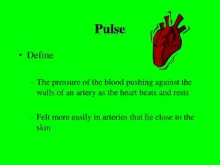
Pulse Oximeter: Function & theory: • Pulse oximetry is the noninvasive measurement of arterial blood oxygen saturation and heart rate . • The Pulse Ox use a spectrophotometric probe . • The pulse oximeter probe contains two light emitting diodes (LEDs) and a photodetector . • the pulse oximeter probe is attached to a highly vascularized tissue, such as a finger or earlobe .
How Pulse Ox Work: • The LEDs emit red and infrared (IR) light . • The signal emitted by the photodetector which convert the red and IR light to volt signal and move it to processor . • The processor compute the percent of blood oxygen saturation (SpO2) according to red and IR absorption and calculate the heart rate . • Finally move the result to display unit .
Absorption of the Lights: • When the light emit to finger it will absorption from : • Tissue . • Bones . • Blood (venous , arterial ) . • After we read the result light signal we can divide it to DC current and AC current as we see in figure
absorption due to pulsatile arterial blood AC absorption due to non pulsatile arterial blood absorption due to venous and capillary blood DC absorption due tissue
Optics and Signal Analysis • SpO2 is a measure of the number of oxygenated hemoglobin molecules in the blood stream relative to the number of molecules of deoxygenated hemoglobin. • Red light is absorbed much by oxygenated hemoglobin than deoxygenated hemoglobin. • IR light is absorbed much by deoxygenated hemoglobin than oxygenated hemoglobin. • The difference in absorption of the two types of light is shown in the figure
Calculate of SpO2 • The AC-signal is isolated by dividing the maximum absorption by the minimum (or DC-signal) value. • the blood oxygen saturation value is a function of the natural logarithm of the maximum and minimum of the red and infrared (IR) signals.
In picture, red refers to the AC-component produced by the pulsing arterial blood, and IR refers to the background DC-component. Max refers to the peak value (amplitude) of the signal, while min refers to the trough (reference) of the signal. • “max” and “min” values in Equation are voltage readings from the photodetector in the pulse oximeter probe.
Pulse Oximeter Probe Components an Function. • The exact wavelengths of the LEDs in the pulse oximeter probe vary among the different probe models and manufacturers. For red LEDs, the wavelength is 660 nm. For infrared LEDs, the wavelengths vary between 880 nm and 950 nm, but 905 nm and 940 nm LEDs are the most common. The probe LEDs emit alternate flashes of red and infrared light at a frequency approximately equal to 400 Hz5. Figure 5 shows a sample output waveform from the LEDs of a pulse oximeter probe.
The photodetector in the pulse oximeter probe is typically a photodiode, which transforms an input signal into a current. The current is then amplified, converted to a voltage, and analyzed by circuitry within the pulse oximeter itself. As the LEDs send alternating pulses of red and infrared light through the tissue and into the photodetector, the photodetector records the corresponding voltages as discrete values and sends them to the pulse oximeter. The pulse oximeter analyzes and compiles these discrete red and infrared voltage values into arrays representing the red and infrared waveforms. The photodetector, therefore, effectively samples the red and infrared waveforms by detecting the alternating red and infrared signals and recording their respective voltage values at a sampling frequency of about 400Hz. Because the signals emitted by the LED are transformed by pulsatile blood flow through the tissue before they are received by the photodetector, the waveforms produced by the voltage measurements are periodic, with a frequency dependent upon heart rate and an amplitude dependent upon oxygen saturation.