Download
1 / 25
310 likes | 916 Views
Structure and function of hemoglobin. Dr. Sumbul Fatma Clinical Chemistry Department of Pathology. Hemoglobin (Hb). A hemeprotein found only in red blood cells Oxygen transport function Contains heme as a prosthetic group Heme reversibly binds to oxygen. The heme group.

E N D
Structure and function of hemoglobin Dr. SumbulFatma Clinical Chemistry Department of Pathology
Hemoglobin (Hb) • A hemeprotein found only in red blood cells • Oxygen transport function • Contains heme as a prosthetic group • Heme reversibly binds to oxygen
The heme group • A complex of protoporphyrin IX and ferrous iron (Fe2+) • Fe2+ is present in the center of heme • Binds to four nitrogens of the porphyrin ring • Plus two additional bonds with: • Histidine residue of globin chain • Oxygen
Page 325 The heme group: Fe2+– porphyrin complex with bound O2
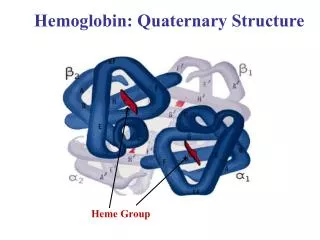
Hemoglobin A (HbA) • Major Hb in adults • Composed of four polypetide chains: • Two α and two β chains • Contains two dimers of αβ subunits • Held together by noncovalent interactions • Each chain is a subunit with a heme group in the center that carries oxygen • A Hb molecule contains 4 heme groups and carries 4 moelcules of O2
T-form of Hb • The deoxy form of Hb • Taut form • The movement of dimers is constrained • Low oxygen affinity form
R-form of Hb • The oxygenated form of Hb • Relaxed form • The dimers have more freedom of movement • High-oxygen-affinity form
Hemoglobin function • Carries oxygen from the lungs to tissues • Carries carbon dioxide from tissues back to the lungs • Normal level: • Males: 14-16 g/dL • Females: 13-15 g/dL
Factors affecting oxygen binding • Three allosteric effectors: • pO2 (partial oxygen pressure) • pH of the environment • pCO2 (partial carbon dioxide pressure) • Availability of 2,3-bisphosphoglycerate
Oxygen Dissociation Curve • The curve is sigmoidal • Indicates cooperation of subunits in O2 binding • Binding of O2 to one heme group increases O2 affinity of others • Heme-heme interaction
P50 (Partial oxygen pressure) • P50 (mm Hg): the pressure at which Hb is 50% saturated with O2 • Indicates affinity of Hb to O2 • High affinity slow unloading of O2 • Low affinity fast unloading of O2 • Lung pO2 is 100 mm Hb saturation 100% • Tissue pO2 is 40 mm Hb saturation reduces to 75% • Oxygen delivery to tissues 25%
The Bohr effect • Effect of pH and pCO2 on: • Oxygenation of Hb in the lungs • Deoxygenation at the tissues • Tissues have lower pH (acidic) than lungs • Due to proton generatation: CO2 + H20 ------> HCO3- + H+ • Protons reduce O2 affinity of Hb • Causing easier O2 release into the tissues • The free Hb binds to two protons
The Bohr Effect • Protons are released and react with HCO3– to form CO2 gas • The proton-poor Hb now has greater affinity for O2 • The Bohr effect removes insoluble CO2 from blood stream • Produces soluble bicarbonate
Availability of 2,3-bisphosphoglycerate • Binds to deoxy-hb and stabilizes the T-form • When oxygen binds to Hb, BPG is released • At high altitudes there is • -increase in no. of RBcs • Increase in conc. Of Hb • Increase in BPG
High altitude and O2 affinity • High altitude decreases Hb O2 affinity • Hypoxia • Increases 2,3 DPG levels • Decreases O2 affinity • Increases O2 delivery to tissues
High O2 affinity • High O2 affinity occurs due to: • Alkalosis • High levels of abnormal Hb (met Hb, CO-Hb) • High levels of Hb F • Multiple transfusion of 2,3 DPG-depleted blood
Fetal Hemoglobin (HbF) • Major hemoglobin found in the fetus and newborn • Tetramer with two α and two g chains • Higher affinity for O2 than HBA • Transfers O2 from maternal to fetal circulation across placenta
HbA2 • Appears ~12 weeks after birth • Constitutes ~2% of total Hb • Composed of two α and two δ globin chains
HbA1c • HbA is slowly and non-enzymatically glycosylated • Glycosylation depends on plasma glucose levels • HbA1c levels are high in patients with diabetes mellitus
Abnormal Hbs • Unable to transport O2 due to abnormal structure • Carboxy-Hb: CO replaces O2 and binds 200X tighter than O2(in smokers) • Met-Hb: Contains oxidized Fe3+ (~2%) that cannot carry O2 • Sulf-HB: Forms due to high sulfur levels in blood (irreversible reaction)