Download
1 / 12
120 likes | 160 Views
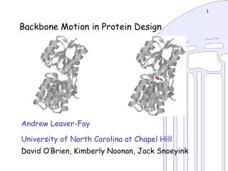
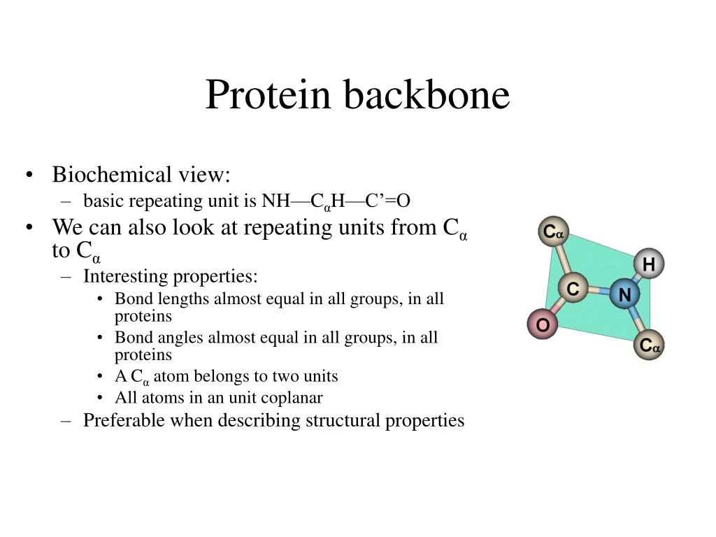
Protein backbone. Biochemical view: basic repeating unit is NH—C α H—C’=O We can also look at repeating units from C α to C α Interesting properties: Bond lengths almost equal in all groups, in all proteins Bond angles almost equal in all groups, in all proteins

E N D
Protein backbone • Biochemical view: • basic repeating unit is NH—CαH—C’=O • We can also look at repeating units from Cα to Cα • Interesting properties: • Bond lengths almost equal in all groups, in all proteins • Bond angles almost equal in all groups, in all proteins • A Cα atom belongs to two units • All atoms in an unit coplanar • Preferable when describing structural properties
Protein backbone • Geometric/Structural view: polypeptide chain divided into • Peptide units • Cα atom and carboxyl group of residue i • Amino group and Cα atom of residue i+1 • Are rigid groups • Rotation on bond C-N is prevented by energy barrier • Peptide units arejoined by covalent bonds between Cα atoms. Thus • Peptides can rotate along 2 bonds: • N-Cα and Cα-C • Two dihedral angles for each unit: Ф (Phi) and Ψ (Psi) • Two degrees of freedom per unit • Determine the conformation of the backbone
Dihedral angles and regular structures • Repeating values of Ф and Ψ along the main chain result in regular structure • repeating values of Ф =-57o and Ψ =-47o give a right-handed helical fold (α-helix) • repetitive values of Ф[-110,-140] and Ψ[+110,+135] give sub chains with conformations that allow interactions between nearby parallel segments (β-sheet) • Most combinations of Ф and Ψ angles are not allowed • Allowed conformations plotted as 2-D chart • Ramachandran plot
Secondary Structure • defined by patterns of hydrogen bonds between backbone amide groups • sidechain-mainchain and sidechain-sidechain hydrogen bonds are irrelevant • The amino acids in the interior/core of a globular protein have hydrophobic side chains • Water soluble proteins fold to pack hydrophobic side chain into interior • Results in hydrophobic core and hydrophilic surface • The main chain must fold into interior, too • Main chain is hydrophilic: • N--H: hydrogen bond donor • C=O: hydrogen bond acceptor • These groups must be neutralized by formation of H bonds secondary structure • Secondary structure • α-helices • β-sheets • form rigid and stable frameworks
α-Helix • Righthanded coiled conformation • backbone N-H group i+4 forms hydrogen bonding with backbone C = O group i • 3.6 residues per turn (5.4 Å, 1.5 Å per residue) • Variations, with chain more loosely or tightly coiled are possible (i+3 or i+5 instead of i+4) but not often • backbone (φ, ψ) dihedral angles around (-60o,-45o) • Sum of φ and ψ angles of consecutive residues about 105o • Has between 4 to 40 residues • All H bonds point in the same direction • Aligned along helical axis • Dipole moments for residues are aligned along axis • Net dipole for α-helix (+ at N-H end and – at C=O end)
β-sheets • Combination of several regions of the chain (not chain adjacent): β-strands • Parallel: all amino acids go in same direction • Evenly spaced H bonds • Antiparallel: amino acids in successive strands alternate directions • Alternate narrowly/widely spaced H bonds • Mixed β-sheet also exist • Have twisted strands: right-handed twist (always) • β-strand: 5 to 10 residues long • Almost fully extended
From secondary structure to structure • Protein structure: built from secondary structures • Connected by loop regions • Various lengths • Irregular shape • Are at the surface of the protein • Reach in charged and polar residues • Easier to predict! • In homologous proteins almost always insertions and deletions occur in the loop regions.
Structure Motifs • Secondary structures connected to form motifs • α-helices and β-sheets in a motif • Adjacent in the 3-dimensional structure • Connected bu loop regions • Combinations of motifs and secondary structures domains
Tertiary structure: • Arrangement of secondary structure • Structural domains • Quaternary structure • More than one polypeptide folded together • Native conformation: direct consequence of • primary structure • chemical environment • water based • oily interior of a cell membrane • So far, no reliable computational method exists to predict the native structure from the amino acid sequence
Structure Classes • Protein structure four classes: • α-domains • core built up only from α-helices • β-domains • core built up only from (usually 2) antiparallel β-sheets • α/β-domains • mostly β-α-β motifs • (mostly) parallel β-sheets surrounded by α-helices • α+β-domains (few cases) • antiparallel β-sheet packed against α-helices