Download
1 / 41
521 likes | 2.11k Views
Aortic Dissection. Jason S. Finkelstein, M.D. Cardiology Fellow Tulane University 8/11/03. Overview. Incidence of aortic dissection is at least 2000 new cases per year Peak incidence is in the sixth to seventh decade Men are affected twice as commonly as women

E N D
Aortic Dissection Jason S. Finkelstein, M.D. Cardiology Fellow Tulane University 8/11/03
Overview • Incidence of aortic dissection is at least 2000 new cases per year • Peak incidence is in the sixth to seventh decade • Men are affected twice as commonly as women • Mortality in the first 48 hours is 1% per hour • Early diagnosis is essential
Pathophysiology • The chief predisposing factor is degeneration of collagen and elastin in the aortic intima media • Blood passes through the tear into the aortic media, separating the media from the intima and creating a false lumen • Dissection can occur both distal and proximal to the tear
Classification • Debakey system • Type I • Originates in the ascending aorta, propagates to the aortic arch and beyond it distally • Type II • Confined to the ascending aorta • Type III • Confined to the descending aorta, and extends distally, or rarely retrograde into the aortic arch
Classification • The Stanford system • Type A • All dissections involving the ascending aorta • Type B • All other dissections regardless of the site of the primary intimal tear • Ascending aortic dissections are twice as common as descending
Predisposing factors • Age, 60-80 yrs old • Long standing history of hypertension • 80% of cases have co-existing HTN • Takayasu’s arteritis • Giant cell arteritis • Syphilis • Collagen disorders • Marfan syndrome (6-9% of aortic dissections) • Ehlers-Danlos syndrome
Other Risk Factors • Congenital Cardiac Anomalies • Bicuspid aortic valve (7-14% of cases) • Coarctation of the aorta • Cocaine • Abrupt HTN, due to catecholamine release • Trauma • Pregnancy (50% of dissections in women <40 yrs) • Iatrogenic (cardiac cath, IABP, cardiac surgery, s/p valve replacement)
Clinical Symptoms • Severe, sharp, “tearing” posterior chest pain or back pain (occurs in 74-90% of pts) • Pain may be associated with syncope, CVA, MI, or CHF • Painless dissection relatively uncommon • Chest pain is more common with Type A dissections • Back or abdominal pain is more common with Type B dissections
Physical Exam • Pulse deficit • Weak or absent carotid, brachial, or femoral pulses • these patients have a higher rate of mortality • Acute Aortic Insufficiency • Diastolic decrescendo murmur • Best heard along the right sternal border
Clinical signs • Acute MI • RCA most commonly involved • Cardiac tamponade • Pleural effusions • Hypertension or hypotension • Hemothorax • Variation in BP between the arms (>30mmHg) • Neurologic deficits • Stroke or decreased consciousness
Clinical Signs • Involvement of the descending aorta • Splanchnic ischemia • Renal insufficiency • Lower extremity ischemia • Spinal cord ischemia
Diagnosis • Generally suspected from the history and PE • In a recent study in 2000, 96% of acute dissection patients could be identified based upon a combination of three clinical features • Immediate onset of chest pain • Mediastinal widening on CXR • A variation in pulse and/or blood pressure (>20 mmHg difference between R & L arm • Incidence >83% when any combination of all three variables occurred
Differential Diagnosis • Acute Coronary Syndrome • Pericarditis • Pulmonary embolus • Pleuritis • Cholecystitis • Perforating ulcer
Diagnostic Tests • EKG • Absence of EKG changes usually helps distinguish dissection from angina • Usually non-specific ST-T wave changes seen • CXR • Cardiac Enzymes
Chest X-Ray • May show widening of the aorta with ascending aorta dissections • Present in 63 % of patients with Type A dissections
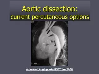
Diagnostic Imaging • Not performed until the patient is medically stable • Has been a dramatic shift from invasive to non-invasive diagnostic strategy • Spiral CT scan • TEE • MRI • Angiography
Imaging • Can identify aortic dissection and other features such as: • Involvement of the ascending aorta • Extent of dissection • Thrombus in the false lumen • Branch vessel or coronary artery involvement • Aortic insufficiency • Pericardial effusion with or without tamponade • Sites of entry and re-entry
Angiography • First definitive test for aortic dissection • Traditionally considered “the gold standard” • Involves injection of contrast media into the aorta • Identifies the site of the dissection • Major branches of the aorta • Communication site between true & false lumen • Can detect thrombus in the false lumen • Disadvantages • Not very practical in critically ill patients • Nephrotoxic contrast • Risks of an invasive procedure
Spiral CT • Sensitivity 83% • Specificity 90 - 100% • Two distinct lumens with a visible intimal flap can be identified • Advantages • Noninvasive • Readily available at most hospitals on an emergency basis • Can differentiate dissection from other causes of aortic widening (tumor, periaortic hematoma, fat) • Disadvantages • Sensitivity lower than TEE and MRI • Intimal flap is seen < 75% of cases • Nephrotoxic contrast is required • Cannot reliably detect AI, or delineate branch vessels
TTE • First used to diagnose aortic dissections in the ’70s • Sensitivity 59-85%, specificity 63-96% • Image quality limited by obesity, lung disease, and chest wall deformities
TEE • Sensitivity 98% Specificity 95% • Advantages • Close proximity of the esophagus to the thoracic aorta • Portable procedure • Yields diagnosis in < 5 minutes • Useful in patients too unstable for MRI • True and false lumens can be identified • Thrombosis, pericardial effusion, AI, and proximal coronary arteries can be readily visualized
TEE • Lower specificity attributed to reverberations atherosclerotic vessels or calcified aortic disease producing echo images that resemble an aortic flap • Disadvantages • Contraindicated in patients with esophageal varices, tumors, or strictures • Potential complications: bradycardia, hypotension, bronchospasm
MRI • Most accurate noninvasive for evaluating the thoracic aorta • Sensitivity 98% • Specificity 98% • Advantages • Safe • Can visualize the whole extent of the aorta in multiple planes • Ability to assess branch vessels, AI, and pericardial effusion • No contrast or radiation • Disadvantages • Not readily available on an emergency basis • Time consuming • Limited applicability in pts with pacemakers or metallic clips
Conclusions • Conventional TTE is of limited diagnostic value in assessment of the thoracic aorta • Both TEE and MRI have excellent sensitivity, however MRI is more specific • MRI is the study of choice for stable patients • TEE is the study of choice for unstable patients
Treatment • Acute dissections involving the ascending aorta are considered surgical emergencies • Dissections confined to the descending aorta are treated medically • Unless patient demonstrates continued hemorrhage into the pleural or retroperitoneal space
Surgical Options • Excision of the intimal tear • Obliteration of entry into the false lumen proximally • Reconstitution of the aorta with interposition of a synthetic vascular graft
Type A Dissections • Operative mortality varies from 7-35% • 27% post-op mortality • Patients who died had a higher rate of in-hospital complications such as strokes, renal failure, limb ischemia, & mesenteric ischemia
Poor prognostic factors • Hypotension or shock • Renal failure • Age> 70 yrs • Pulse deficit • Prior MI • Underlying pulmonary disease • Preoperative neurologic impairment • Renal and/or visceral ischemia • Abnormal EKG, particularly ST elevation
Medical therapy • Reduce systolic BP to 100 to 120 mmHg or the lowest level that is tolerated • IV Beta blockers • Propanolol (1-10 mg load, 3mg/hr) • Labetalol (20 mg bolus, 0.5 to 2 mg/min) • If SBP remains >100mmHg, nitroprusside should be added • Do not use without beta blockade • Avoid hydralazine • Surgical intervention for Type B dissections reserved for patients with a complicated course
Long Term Outcome • Type A • Survival at 5 yrs – 68% • Survival at 10 yrs – 52 % • Type B • 5 yrs – 60 - 80% • 10 yrs – 40 – 80% • Spontaneous healing of dissection is uncommon
Long-Term Management • Medical therapy • Oral Beta-blockers (reduces aortic wall stress) • Keep BP < 135/80 mmHg (combination therapy) • Avoidance of strenuous physical activity • Serial imaging • Thoracic MR scan prior to discharge • f/u scans at 3, 6, and 12 months • Subsequent screening studies done every 1-2 yrs if no evidence of progression