Download
1 / 15
330 likes | 1.14k Views
Aortic Dissection. Aortic Dissection. From: iradonline.org/images/aorta-layers.gif. Types of Aortic Dissections. Stanford Classification Type A = involves ascending aorta Type B = does not involve ascending aorta DeBakey Classification Type I = ascending and descending aorta

E N D
Aortic Dissection From: iradonline.org/images/aorta-layers.gif
Types of Aortic Dissections • Stanford Classification • Type A = involves ascending aorta • Type B = does not involve ascending aorta • DeBakey Classification • Type I = ascending and descending aorta • Type II = ascending aorta only • Type III = descending aorta only
From: www.massgeneral.org/tac/patients/diseases.asp?id=a_dissection
Aortic Dissection Risk Factors Typically: • Systemic Hypertension • Systemic Hypertension • Did I mention systemic hypertension? • Present in 60-90% of patients In younger patients, must consider other factors: • Bicuspid aortic valve (9% under 40 in one review) • Inflammatory disease (giant cell, syphilitic aortitis, RA, etc.) • Collagen Diseases (ED Syndrome, Marfan’s (50% in those under 40 in one review)) • Preexisting aortic aneurysm • Coarctation Others: • CABG • Trauma • Iatrogenic (intravascular procedures) • Cocaine use (thought to be catecholamine mediated)
Clinical Manifestations/Findings • Acute onset of tearing chest pain, frequently radiating to the back • Syncope • Asymmetric BP in UE • Weak/absent peripheral pulses • Aortic Insufficiency/Heart Failure • MI • Renal failure • Paraplegia • Back Pain • Pericardial effusion
Many different presentations; how to diagnose clinically? • One relatively small study found that in 250 patients with acute chest and/or back pain, certain findings were particularly relevant: sudden onset of tearing/ripping chest pain, widening of mediastinum or aorta (or both), and pulse or BP differentials (or both). • Absense of all three, low probability (7%) • Characteristic chest pain, intermediate probability (31%) • Mediastinal Widening, intermediate probability (39%) • Pulse/BP differential, high probability (>83%) • Combination of all three, high probability (>83%)
Bottom line is that you need imaging to assist in the diagnosis
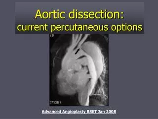
Imaging Studies • CXR: abnormal in majority of patients (90% sensitive), but cannot rule out dissection (although completely normal imaging is helpful) • CT: up to 98% sensitive and up to 100% specific (depending on the study) • TEE: up to 98% sensitive 95% specific • MRI: sensitivity & specificity >98% • Aortography: sensitivity <90%
Acute Management • Risk of death for untreated acute dissection is estimated at 1% per hour • IV antihypertensives (goal of SBP <120 or lower if tolerated) and negative ionotropic agents (goal HR < 60) • Pain control • Cardiac monitoring, preparation for aggressive resuscitation • Ultimate goal initially is to prevent death and irreversible end-organ damage
Definitive Therapy • Ascending Dissections Surgery ASAP • Descending Dissections • If stable and uncomplicated: managed medically • If unstable or complications: surgery
Subsequent Management • Blood pressure control… the lower, the better • Serial imaging with MRI/CT on annual basis to evaluate for aneurysm formation, anastomotic leakage, recurrent dissection • Consideration of second operation to repair aforementioned complications