Download
1 / 42
500 likes | 1.82k Views
Page 1069. Figure 28-1 The biosynthetic origins of purine ring atoms. Page 1071. Figure 28-2 The metabolic pathway for the de novo biosynthesis of IMP. Page 1072. Figure 28-3 The proposed mechanism of formylglycinamide ribotide (FGAM) synthetase. Page 1074.

E N D
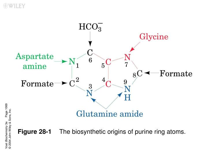
Page 1069 Voet Biochemistry 3e © 2004 John Wiley & Sons, Inc. Figure 28-1 The biosynthetic origins of purine ring atoms.
Page 1071 Voet Biochemistry 3e © 2004 John Wiley & Sons, Inc. Figure 28-2 The metabolic pathway for the de novo biosynthesis of IMP.
Page 1072 Voet Biochemistry 3e © 2004 John Wiley & Sons, Inc. Figure 28-3 The proposed mechanism of formylglycinamide ribotide (FGAM) synthetase.
Page 1074 Voet Biochemistry 3e © 2004 John Wiley & Sons, Inc. Figure 28-4 IMP is converted to AMP or GMP in separate two-reaction pathways.
Page 1075 Voet Biochemistry 3e © 2004 John Wiley & Sons, Inc. Figure 28-5 Control network for the purine biosynthesis pathway.
Page 1077 Voet Biochemistry 3e © 2004 John Wiley & Sons, Inc. Figure 28-6 The biosynthetic origins of pyrimidine ring atoms.
Page 1077 Voet Biochemistry 3e © 2004 John Wiley & Sons, Inc. Figure 28-7 Metabolic pathway for the de novo synthesis of UMP.
Page 1078 Voet Biochemistry 3e © 2004 John Wiley & Sons, Inc. Figure 28-8 Reactions catalyzed by eukaryotic dihydroorotate dehydrogenase.
Page 1079 Voet Biochemistry 3e © 2004 John Wiley & Sons, Inc. Figure 28-9 Proposed catalytic mechanism for OMP decarboxylase.
Page 1080 Voet Biochemistry 3e © 2004 John Wiley & Sons, Inc. Figure 28-10 Synthesis of CTP from UTP.
Page 1080 Voet Biochemistry 3e © 2004 John Wiley & Sons, Inc. Figure 28-11 Regulation of pyrimidine biosynthesis. The control networks are shown for (a) E. coli and (b) animals.
Page 1082 Voet Biochemistry 3e © 2004 John Wiley & Sons, Inc. Figure 28-12a Class I ribonucleotide reductase from E. coli. (a) A schematic diagram of its quaternary structure.
Page 1082 Voet Biochemistry 3e © 2004 John Wiley & Sons, Inc. Figure 28-12bClass I ribonucleotide reductase from E. coli. (b) The X-ray structure of R22.
Page 1082 Voet Biochemistry 3e © 2004 John Wiley & Sons, Inc. Figure 28-12c Class I ribonucleotide reductase from E. coli. (c) The binuclear Fe(III) complex of R2.
Page 1082 Voet Biochemistry 3e © 2004 John Wiley & Sons, Inc. Figure 28-12d Class I ribonucleotide reductase from E. coli. (d) The X-ray structure of the R1 dimer.
Page 1083 Voet Biochemistry 3e © 2004 John Wiley & Sons, Inc. Figure 28-13 Enzymatic mechanism of ribonucleotide reductase.
Page 1085 Voet Biochemistry 3e © 2004 John Wiley & Sons, Inc. Figure 28-14a Ribonucleotide reductase regulation. (a) A model for the allosteric regulation of Class I RNR via its oligomerization.
Page 1085 Voet Biochemistry 3e © 2004 John Wiley & Sons, Inc. Figure 28-14b Ribonucleotide reductase regulation. (b) The X-ray structure of the R1 hexamer, which has D3 symmetry, in complex with ADPNP as viewed along its 3-fold axis.
Page 1085 Voet Biochemistry 3e © 2004 John Wiley & Sons, Inc. Figure 28-14c Ribonucleotide reductase regulation. (c) The R1·ADPNP hexamer as viewed along the vertical 2-fold axis in Part b.
Page 1086 Voet Biochemistry 3e © 2004 John Wiley & Sons, Inc. Figure 28-15 X-Ray structure of human thioredoxin in its reduced (sulfhydryl) state.
Page 1087 Voet Biochemistry 3e © 2004 John Wiley & Sons, Inc. Figure 28-16 Electron-transfer pathway for nucleoside diphosphate (NDP) reduction.
Page 1087 Voet Biochemistry 3e © 2004 John Wiley & Sons, Inc. Figure 28-17a X-Ray structures of E. coli thioredoxin reductase (TrxR). (a) The C138S mutant TrxR in complex with NADP+.
Page 1087 Voet Biochemistry 3e © 2004 John Wiley & Sons, Inc. Figure 28-17b The C135S mutant thioredoxin reductase (TrxR) in complex with AADP+, disulfide-linked to the C35S mutant of Trx.
Page 1089 Voet Biochemistry 3e © 2004 John Wiley & Sons, Inc. Figure 28-18a X-Ray structure of human dUTPase. (a) The molecular surface at the substrate binding site showing how the enzyme differentiates uracil from thymine.
Page 1089 Voet Biochemistry 3e © 2004 John Wiley & Sons, Inc. Figure 28-18b X-Ray structure of human dUTPase. (b) The substrate binding site indicating how the enzyme differentiates uracil from cytosine and 2-deoxyribose from ribose.
Page 1090 Voet Biochemistry 3e © 2004 John Wiley & Sons, Inc. Figure 28-19 Catalytic mechanism of thymidylate synthase.
Page 1091 Voet Biochemistry 3e © 2004 John Wiley & Sons, Inc. Figure 28-20 The X-ray structure of the E. coli thymidylate synthase–FdUMP–THF ternary complex.
Page 1091 Voet Biochemistry 3e © 2004 John Wiley & Sons, Inc. Figure 28-21 Regeneration of N5,N10-methylenetetrahydrofolate.
Page 1091 Voet Biochemistry 3e © 2004 John Wiley & Sons, Inc. Figure 28-22 Ribbon diagram of human dihydrofolate reductase in complex with folate.
Page 1093 Voet Biochemistry 3e © 2004 John Wiley & Sons, Inc. Figure 28-23 Major pathways of purine catabolism in animals.
Page 1094 Voet Biochemistry 3e © 2004 John Wiley & Sons, Inc. Figure 28-24a Structure and mechanism of adenosine deaminase. (a) A ribbon diagram of murine adenosine deaminase in complex with its transition state analog HDPR.
Page 1094 Voet Biochemistry 3e © 2004 John Wiley & Sons, Inc. Figure 28-24b Structure and mechanism of adenosine deaminase. (b) The proposed catalytic mechanism of adenosine deaminase.
Page 1095 Voet Biochemistry 3e © 2004 John Wiley & Sons, Inc. Figure 28-25 The purine nucleotide cycle.
Page 1095 Voet Biochemistry 3e © 2004 John Wiley & Sons, Inc. Figure 28-26a X-Ray structure of xanthine oxidase from cow’s milk in complex with salicylic acid. (a) Ribbon diagram of its 1332-residue subunit.
Page 1095 Voet Biochemistry 3e © 2004 John Wiley & Sons, Inc. Figure 28-26b X-Ray structure of xanthine oxidase from cow’s milk in complex with salicylic acid. (b) The enzyme’s redox cofactors and salicylic acid (Sal).
Page 1096 Voet Biochemistry 3e © 2004 John Wiley & Sons, Inc. Figure 28-27 Mechanism of xanthine oxidase.
Page 1097 Voet Biochemistry 3e © 2004 John Wiley & Sons, Inc. Figure 28-28 Degradation of uric acid to ammonia.
Page 1097 Voet Biochemistry 3e © 2004 John Wiley & Sons, Inc. Figure 28-29The Gout, a cartoon by James Gilroy (1799).
Page 1098 Voet Biochemistry 3e © 2004 John Wiley & Sons, Inc. Figure 28-30 Major pathways of pyrimidine catabolism in animals.
Page 1099 Voet Biochemistry 3e © 2004 John Wiley & Sons, Inc. Figure 28-31 Pathways for the biosynthesis of NAD+ and NADP+.
Page 1100 Voet Biochemistry 3e © 2004 John Wiley & Sons, Inc. Figure 28-32 Biosynthesis of FMN and FAD from the vitamin precursor riboflavin.
Page 1101 Voet Biochemistry 3e © 2004 John Wiley & Sons, Inc. Figure 28-33 Biosynthesis of coenzyme A from pantothenate, its vitamin precursor.