Download
1 / 2
30 likes | 558 Views
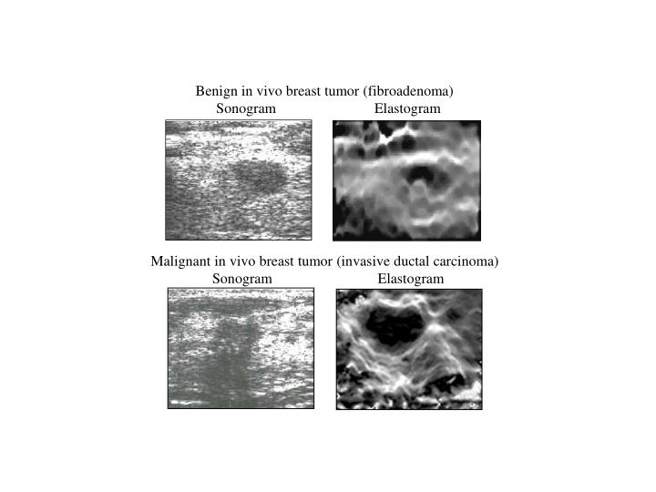
Benign in vivo breast tumor (fibroadenoma) Sonogram Elastogram. Malignant in vivo breast tumor (invasive ductal carcinoma) Sonogram Elastogram.

E N D
Benign in vivo breast tumor (fibroadenoma) Sonogram Elastogram Malignant in vivo breast tumor (invasive ductal carcinoma) Sonogram Elastogram
Sonograms and corresponding elastograms of benign and malignant tumors of the female breast in vivo. The elastograms were generated using a compression of approximately 1%. Black denotes stiffer and white denotes softer tissue, respectively. Not only do elastograms provide for better detection of both tumors but also comparison of the two types of images leads to possible differentiation between benign and malignant. Benign fibroadenomas generally have smooth regular borders and their measured transverse diameter on elastograms is almost always the same or smaller than their diameter on sonography (upper panel). The transverse diameter of cancers is generally larger on elastograms than on sonograms (lower panel). This is most likely due to the firm desmoplastic reaction surrounding cancers being included as part of the tumor measurement in elastography. Using the combination of tumor measured relative stiffness and the difference between the sonographic and elastographic tumor measured diameter, the differentiation of benign versus malignant tumors has been shown possible. (Image courtesy of Elisa Konofagou, Brian Garra and Jonathan Ophir, University of Texas Medical School – Houston and University of Vermont).