Download
1 / 42
460 likes | 1.85k Views
Blood Cells Simon Hunt Dunn School of Pathology http://users.path.ox.ac.uk/~svhunt/internal/bloodcells OR http://www.weblearn.ox.ac.uk/bodington/site/medsci/undergrad/med/teach/lectsupp/hunt/ Images from Wheater’s Functional Histology, 4 th edn

E N D
Blood Cells Simon Hunt Dunn School of Pathology http://users.path.ox.ac.uk/~svhunt/internal/bloodcells OR http://www.weblearn.ox.ac.uk/bodington/site/medsci/undergrad/med/teach/lectsupp/hunt/ Images from Wheater’s Functional Histology, 4th edn Some electron micrographs from the collection of the late Drs Poole and French, Dunn School
Purposes of This Presentation • How structure relates to function in erythrocytes (red blood cells) • To the level of syllabus section 5.4.1 • As living and dying ultra-specialised (“differentiated”) cells • Ditto for leukocytes (white blood cells) • To the level of syllabus section 5.4.2 • A functional “Who’s Who?” • How we find out about the physiological properties of blood cells • Illustrated by some disease conditions Blood Cells
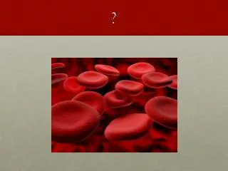
Haematocrit tube ~ 45% Red and white blood cells Blood Cells
7.2mm Erythrocytes – Greek: “Red Hollows” • Pink stain – eosin, not haem • Haemoglobin is a basic protein • Binds acid dyes (e.g. eosin) • Scanning e-m • Always black & white Blood Cells
Blood cells, computer-enhanced © Dennis Kunkel Microscopy, Inc http://www.denniskunkel.com Blood Cells
Erythrocytes can form Rouleau (pl. Rouleaux) • Like a stack of coins • Only when blood flow is slow http://www.finchcms.edu/cms/anatomy/histology/histology/blood/blood.html#blood Blood Cells
Rouleaux 2 • by fluorescence • Red shows DNA stain of leukocytes • Shows that rbc have no nuclei http://www.cyto.purdue.edu/flowcyt/educate/photos/cells/images.htm Blood Cells
(a):discocyte, (b) stomatocyte and (c) echinocyte from Harriet Gershon www.els.net Erythrocyte shape • Anucleate (no nucleus) • also, no mitochondria • Shape depends on water content • Osmotic effects of solutes, especially ions • Shape maintained by cytoskeleton • Spectrin, ankyrin, & other membrane proteins Blood Cells
Advantages of being anucleate • Better surface-volume ratio • About 25% greater than if an equivalent sphere • Improves gas exchange • Improves deformability • To fit through capillaries • diameter of true capillaries is only 5-10 mm, often less • Less work for heart as a pump • Pumps approximately 3 kg of erythrocytes per minute • 40% of total mass would be nucleus • Saves pumping 1 to 1.5 tons per day Blood Cells
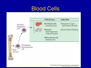
Disadvantages of being anucleate • No further protein synthesis or repair • Finite cell lifespan • 120 days on average • Requires vast new replacement cell production • Red blood cell production = “erythropoiesis” • Equally true for blood platelets • Platelet production = “thrombopoiesis” Blood Cells
Consequences of lacking mitochondria • Have to survive on anaerobic metabolism (glycolysis) • Energy needs not great, mainly ion pumps • Depends solely on blood glucose for energy supply • the glycolytic intermediate, 2,3-bisphosphoglycerate (2,3-BPG), is produced by an erythrocyte enzyme • BPG shifts dissociation curve to unload O2 from HbO2 Blood Cells
ErythrocyteContents 1 • Haemoglobin ~750 gm per adult body • Globin protein, alpha2beta2,~650 million molecules per cell • Haem prosthetic group • One Fe2+ per haem, ~2.2 gm per whole body • About 2/3 of all body iron • Must not oxidise to Fe3+ ( “methaemoglobin”) • cell needs reducing conditions • Functions in O2 and CO2 transport • Dr Dorrington’s lectures later this term Blood Cells
ErythrocyteContents 2 • Glucose metabolising enzymes • Anaerobic glycolysis (Embden-Meyerhof Pathway) • Pentose Phosphate shunt • Uses G6PDH, Glucose-6-Phosphate Dehydrogenase, an X-linked enzyme • generates NADPH, slows build-up of oxidised proteins associated with erythrocyte ageing • thus maintains Glutathione, a Cysteine-containing tripeptide, in the reduced state G6PDH deficiency is revealed by a serious haemolytic crisis when broad beans or the (now-obsolete) anti-malarial Pamaquine are ingested Blood Cells
[Na+] = 6 mM [K+] = ~100 mM ErythrocyteContents 3 • Ions, especially K+ • Maintained by membrane-associated ATP-dependent Na+-K+ ion exchanger PLASMA concns: [Na+] = 140 mM [K+] = 3.5 - 5 mM Blood Cells
Erythrocyte membrane – “ghosts” Blood Cells
Membrane protein functions • Cytoskeletal proteins maintain shape • spectrin, ankyrin, Band III • Channels, pores or pumps • Cations, anions, water, glucose • Glyoproteins & glycolipids: display extracellular carbohydrate • glycophorins maintain net negative charge • blood group substances • Regulatory proteins • complement-absorbing components • anti-inflammatory action Blood Cells
Osmotic effects “Crenated” From http://arbl.cvmbs.colostate.edu/hbooks/cmb/cells/pmemb/osmosis.html Blood Cells
http://www.physiology.rwth-aachen.de/user/martin/DuUndDeinBlut/Bl-Terminologie-d.htmlhttp://www.physiology.rwth-aachen.de/user/martin/DuUndDeinBlut/Bl-Terminologie-d.html Hypotonic haemolysis • “Hypo-” means lower than normal • i.e. solute concentration outside is less than inside cell • allow for ions; count all osmotically active particles; use “Osmolarity” • water potential outside cell > intracellular • Cell membrane semi-permeable • Water permeates via “Aquaporin” proteins • >250 times cell volume crosses membrane per second Blood Cells
Normal http://hsc.virginia.edu/medicine/clinical/pathology/educ/innes/text/rcd/membrane.html Diseased - fragile Osmotic fragility test Blood Cells
DHAG e-m#328 Erythrocytes can deform • To squeeze through arterioles or capillaries • Tend to keep to central axis of vessel • Plasma-rich at circumference • Blood is “visco-elastic” • Not easy to find good artificial substitutes NB rbc are electron-dense – why? Blood Cells
Erythrocytes can deform 2 Blood Cells
Rheology - the flow properties of blood suspensions • The Fåhraeus-Lindqvist effect(small-diameter phenomenon) • rbc concentration is lower near the vessel wall and higher in the centre • hence vessel haematocrit decreases in the smaller branches of the vasculature i.e. more fluid and fewer cells in capillaries • hence get misleading blood cell concentrations if capillaries are sampled Blood Cells
Anomalous viscosity of blood • Viscosity of blood increases with decreased velocity • Blood flow is low in small vessels (1 mm/sec) – • viscosity can increase 10 times just because of slow velocity • due to adherence of RBCs to each other (form rouleaux) and to vessel walls • shear forces no longer enough to deform RBC, so they appear more rigid • Effect is even more noticeable • if membrane more rigid, e.g. Spectrin defect, or becomes crenated • in aged erythrocytes • if there are inclusions inside cells, e.g. sickled cells • if rbs are enlarged osmotically • In what circumstances might this matter clinically? Blood Cells
Spectrin defect Ankyrin defect Band III defect Spectrin deficiency Decreased rbc deformability, osmotic fragility Splenic conditioning: further loss of membrane surface area Rbc entrapment in splenic cords Macrophage removal of severely abnormal rbc http://hsc.virginia.edu/medicine/clinical/pathology/educ/innes/text/rcd/membrane.html Hereditary spherocytosis 1 Loss of membrane surface area micro-spherocytosis Blood Cells
Hypersplenism in hereditary spherocytosis Blood Cells
Summary - erythrocytes • Vast numbers, steady turnover • No nucleus, no mitochondria • Glucose essential as energy source • To maintain reducing conditions, and for ion pumps • Cells become “aged” as oxidation products build up • Lipid bilayer membrane, with attached & inserted glycoproteins • Cytoskeleton • Biconcave disc, but deformable • Flexibility important for proper plasma flow • Semi-permeable properties lysis if not in isotonic medium • Will examine erythropoiesis in next lecture Blood Cells
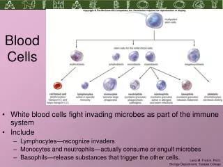
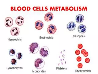
granulocytes Mononuclear cells Leukocytes – Greek: “White Hollows” Blood Cells
Neutrophils 1 Blood Cells
Neutrophils 2 • The most abundant leukocyte in blood • Filled with granules • Lysosomes fuse with ingested phagosome • Secrete toxic chemicals • Very active migration • Sensitive to chemotactic factors which attract them to infection site Blood Cells
Neutrophils: functions as effector cells • Synonyms • Polymorphonuclear leukocytes, = PMNs or “Polys” • Don’t stain strongly with either eosin or basic dyes • Raised numbers (“neutrophilia” during acute bacterial infections • Increased mobilisation from extensive reserves • Increased production from progenitors • Adhere to vessel walls and transmigrate to areas of infection in tissues (acute inflammation) • Engulf bacteria, kill rapidly with very toxic molecules (incl strong oxidisers comparable to bleach) • Collateral damage to host cells, plus dead bugs, pus • Defects in adhesion molecules, or in killing mechanism, serious pyogenic (pus-forming) infections Blood Cells
Eosinophils • Generally larger than neutrophils • Stain orange-pink with eosin • Contain abundant basic protein • Elevated levels in • tropical parasite infections • defence against single-celled and multicellular parasites • Chronic allergic conditions • May reduce hypersensitivity via histaminase Blood Cells
Eosinophil e-m • Large ovoid granules, very electron dense • Actively phagocytic • Passively adsorb certain kinds of antibodies • helps them recognise targets • Can exocytose (spit out) toxic substances • Different chemicals from neutrophils Blood Cells
Basophil • Least common leukocyte • Stains with basic dyes • Precursor of mast cells in tissues • Mast cells release histamine etc in allergies Blood Cells
Monocyte • ~5 – 10% of wbcs • Nucleus often kidney-shaped • No obvious granules • Precursor of macrophages in tissues • Macro = “big”; phage = “eat” Blood Cells
Lymphocyte • Next most common after neutrophils • No obvious granules • Except for Natural Killer subset • Subsets • T, B Blood Cells
Autoradiograph by J.L. Gowans Lymphocytes • Vary in size • Small = very dormant, out of cycle, long-lived • After stimulation memory cells • Large = rapidly dividing • Incorporate DNA synthesis precursors • “Lymphoblasts” • Intermediates before full maturation • To antibody-forming cells in tissues (very rare in blood) • To cytotoxic T lymphocytes • These are from lymph (hence no rbc) Blood Cells
Lymphocytes: small and large by e-m Blood Cells
Lymphocytes patrol continuously • To connect functionally all the dispersed lymphoid tissues • Blood lymph blood ……. • Leave blood at special endothelium only on certain venules, within lymph nodes, tonsils & other lymphoid organs • Ensures rare clones with a given specificity have a good chance of encountering their specific stimulus http://www.geocities.com/CapeCanaveral/Hangar/1962/page3.html Blood Cells
Lymphocytes emigrate via High Endothelial Venules • To seek antigens outside the blood stream, which have been filtered, processed and presented by cells in lymphoid tissues Blood Cells
Summary of leukocytes 1 • Granulocytes include: • Neutrophils • Move promptly into tissues to deal with any noxious event harmful to the body: infection, tissue damage and so on • Engulf, kill and digest invaders inflammation and perhaps pus • Eosinophils • Defence against some parasites, in collaboration with antibodies • May diminish some immediate-type hypersensitivity reactions • Basophils • Move into tissues to become mast cells Blood Cells
Summary of leukocytes 2 • Monocytes • Precursors of tissue macrophages, slower to act than neutrophils • Lymphocytes • Each call has one specificity for antigen, therefore needs to continuously patrol to meet up with the right molecule • perpetual motion into and out of vasculature - “lymphocyte recirculation” Blood Cells
Reading and online materials • Wheater’s Functional Histology, 4th edn, chapter 3 • http://www-micro.msb.le.ac.uk/MBChB/2a.html • Leicester, immune system cells only, not complete haematology • http://www.medinfo.ufl.edu/year1/histo/review/lab11.html • Florida, in-depth, with self-test Qs at: http://www.medinfo.ufl.edu/year1/histo/quiz/mh10.html • http://www.ultranet.com/~jkimball/BiologyPages/B/Blood.html#RBCs • (textbook account, excellent hyperlinked glossary) • http://www.aum.iawf.unibe.ch/HemoSurf/Demo_E/Gallery/NRB_fr.htm • (proper haematologist’s site) Blood Cells