Download
1 / 21
240 likes | 543 Views
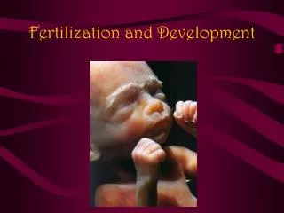
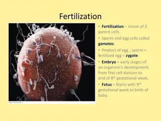
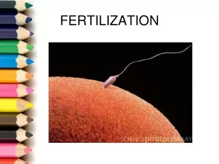
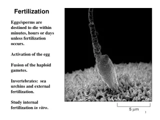
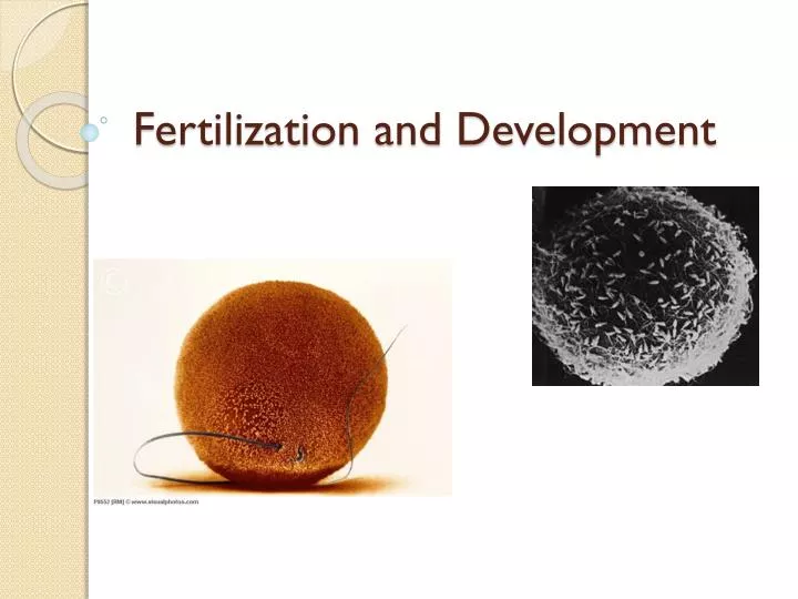
Fertilization and Development. Fertilization. Until this point, we have assumed that the released egg was not fertilized by a sperm However, if sperm are in the Fallopian tube, there is a chance that the egg will become fertilized Sperm attaches by finding binding sites on egg

E N D
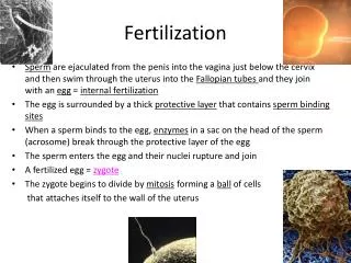
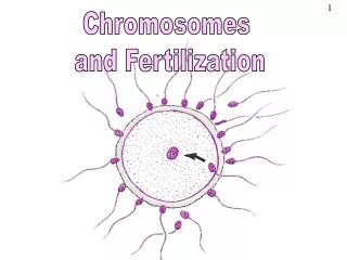
Fertilization • Until this point, we have assumed that the released egg was not fertilized by a sperm • However, if sperm are in the Fallopian tube, there is a chance that the egg will become fertilized • Sperm attaches by finding binding sites on egg • Layer containing sites then dissolves, removing possibility of polyspermy
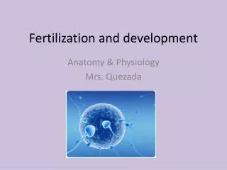
Two haploid nuclei merge to form diploid zygote • Will begin to develop even before it reaches the uterus
Early Development • Divided into three stages: • Implantation • Gastrulation • Neurulation
Neurulation • After fertilization, zygote goes through a series of mitotic divisions, with very little time between each • Reaches around 64 cells (solid ball), called a morula • Morula is not much larger than original single cell • Begins to hallow out, becomes blatocyst
Blastocyst will secrete digestive enzymes to burrow into uterine lining (recall it should be thickest now) about one week after fertilization • This is the implantation • Can cause spotting for the female
Cluster of cells form inside blastocyst • This inner cell mass will become the embryo • Trophoblast surrounding this will become the placenta and umbilical cord
Gastrulation • ICM differentiates into three layers • Ecotoderm – skin and nervous system • Endoderm – lining of digestive track and internal organs • Mesoderm – inner tissue and organs
Amniotic cavity Primitive streak Mesoderm Ectoderm Endoderm Primitive streak is where cell grow out of to form mesoderm
Neurulation • Formation of the CNS • Block of mesoderm differentiates into notochord – eventually becoming the spine
As the notochord develops, the neural groove changes shape, producing neural folds Neural crest Neural fold Notochord
Gradually, these folds move together to create a neural tube from which the spinal cord and the nervous system develop Neural tube Neural crest Ectoderm Notochord
Extraembryonic Membranes • Trophoblast and mesoderm continue to develop, forming two membranes called amnion and the chorion • The amnion develops into a fluid-filled amniotic sac, which cushions and protects the developing embryo • Chorion forms finger-like projections, connecting mother to fetus
Amniotic sac Placenta Umbilical cord Uterus Amnion Fetus
Maternal portion of placenta Fetal portion of placenta Chorionic villus Amnion Umbilical cord Maternal artery Umbilical arteries Maternal vein Umbilical vein • The chorionic villi and uterine lining form the placenta
The Placenta • Serves as embryo’s organ for respiration, nourishment, and excretion • Protects fetus from many, but not all, harmful substances • Viruses such as HIV and substances such as alcohol pass through
First Trimester • Embryo becomes fetus at eight weeks • Most major organs and tissues formed • Umbilical chord develops, connects baby to mother
Second Trimester • The heart can be heard with a stethoscope • Bone replaces cartilage that forms the early skeleton • A layer of soft hair grows over the fetus’s skin • The fetus grows and the mother can feel it moving
Third Trimester • The fetus doubles in mass • It can now regulate its body temperature • The central nervous system and lungs completely develop • Gestation
Childbirth • Mother’s pituitary gland secretes oxytocin • Causes rhythmic, involuntary contractions of muscles around uterine wall • This is known as labour • Amniotic sac will break (water breaking) • Opening to cervix expands, convulsions push baby out through vagina • The Birth Video!