Download
1 / 21
220 likes | 358 Views
Fertilization and Development. Human Anatomy and Physiology. Fertilization. Fusion of sperm and an ovum Occurs in the fallopian tubes of the female An oocyte is viable for 24 hours; sperm are viable for 72 hours.

E N D
Fertilization and Development Human Anatomy and Physiology
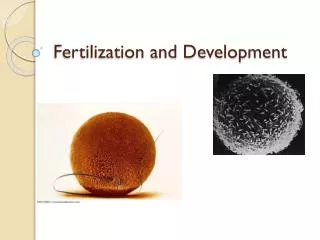
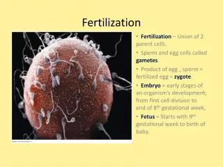
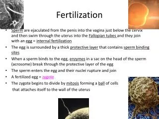
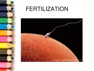
Fertilization • Fusion of sperm and an ovum • Occurs in the fallopian tubes of the female • An oocyte is viable for 24 hours; sperm are viable for 72 hours. • Takes sperm 1-2 hours to complete journey up the female duct system. Must swim against the cilia current.
Fertilization and Implantation Fallopian tube Zygote 4 cells 2 cells Morula Blastocyst Uterine wall Ovary Section 39-4 Day 2 Day 3 Day 1 Day 4 Fertilization Day 0 Day 7 Implantation of blastocyst Egg released by ovary
Cleavage • A rapid series of mitotic divisions • Begins with zygote and ends with a blastocyst
Implantation Blastocyst implanted into endometrium of uterus
LE 46-15a Cleavage starts Cleavage continues Ovary Fertilization occurs The blastocyst implants Uterus Ovulation Endometrium From ovulation to implantation
Pregnancy • Placenta • Formed when the outer layer of the blastocyst mingles with the endometrium • Connects to fetus by the umbilical cord • Serves as respiratory, nutritive, and excretory needs of the embryo • Produces hormones of pregnancy including estrogen and progesterone • Functioning by 3rd week • The tissue of the fetus releases human chorionic gonadotropin (HCG), which helps maintain the uterine lining.
LE 46-16 Maternal veins Maternal arteries Placenta Maternal portion of placenta Umbilical cord Chorionic villus containing fetal capillaries Fetal portion of placenta (chorion) Maternal blood pools Umbilical arteries Uterus Fetal arteriole Umbilical vein Fetal venule Umbilical cord Placental circulation. From the fourth week of development until birth, the placenta, a combination of maternal and embryonic tissues, transports nutrients, respiratory gases, and wastes between embryo or fetus and the mother. Maternal blood enters the placenta in arteries, flows through blood pools in the endometrium, and leaves via veins. Embryonic or fetal blood which remain in vessels enters the placenta through arteries and passes through capillaries in chorionic villi, where oxygen and nutrients are acquired. Fetal blood leaves the placenta through veins leading back to the fetus.
20 weeks. 14 weeks. 5 weeks.
Pregnancy: First Trimester • Radical change for the mother and the embryo • During its first 2 to 4 weeks, the embryo obtains nutrients directly from the endometrium until placenta forms • Blood from the embryo travels to the placenta through arteries of the umbilical cord and returns via the umbilical vein • Main period of organogenesis • development of the body organs • Heart develops first • Neural tube develops • All body systems appear by Week 8 – Now a Fetus
Pregnancy: Second Trimester • During the second trimester • The fetus grows and is very active • The mother may feel fetal movements • The uterus grows enough for the pregnancy to become obvious
Pregnancy: 3rd Trimester • More growth • Kicking, rolling, stretching • Eyes open – Week 32 • Lungs mature • Rotates to head-down position
Childbirth • Due Date is 280 days from the start of the last menstrual period
Initiation of Labor • Estrogen reaches highest level of pregnancy causing progesterone to stop quieting the uterine muscles • Leads to irregular contractions called Braxton’s Hicks (false labor) • Two more chemical signals convert false labor into real labor pains • Oxytocin • Prostaglandins
So…. • Oxytocin and prostaglandins initiate the rhythmic, expulsive contractions of true labor • Positive feedback mechanism • stronger contractions lead to release of more oxytocin leading to even stronger contractions
LE 46-18 Estrogen Oxytocin from ovaries from fetus and mother’s posterior pituitary Induces oxytocin receptors on uterus Positive feedback Stimulates uterus to contract Stimulates placenta to make Prostaglandins Stimulate more contractions of uterus
Stages of Labor • Dilation Stage • Cervix fully dilated by baby’s head to 10 cm. • Cervix softens • Water breaks (amniotic fluid released) • Lasts 6-12 hours • Expulsion Stage • Time from full dilation to delivery of baby • Can take as long as 2 hours, typically 50 minutes (20 minutes if not first child) • Placental Stage • Delivery of placenta • Takes 15 minutes
Placenta Uterus Umbilical cord Placenta (detaching) Uterus Cervix Umbilical cord Delivery of the placenta Expulsion: delivery of the infant Dilation of the cervix
Contraception and Abortion • Contraception, the deliberate prevention of pregnancy, can be achieved in a number of ways • Some general contraceptive methods: • Preventing release of eggs and sperm • Keeping sperm and egg apart • Preventing implantation of an embryo • Abortion is the termination of a pregnancy
LE 46-20 Male Female Event Event Method Method Production of viable oocytes Production of viable sperm Vasectomy Combination birth control pill (or injection, patch, or vaginal ring) Ovulation Sperm transport down male duct system Abstinence Abstinence Condom Coitus interruptus (very high failure rate) Sperm deposited in vagina Capture of the oocyte by the oviduct Tubal ligation Spermicides; diaphragm; cervical cap; progestin alone (minipill, implant, or injection) Sperm movement through female reproductive tract Transport of oocyte in oviduct Meeting of sperm and oocyte in oviduct Morning-after pill (MAP) Union of sperm and egg Progestin alone Implantation of blastocyst in properly prepared endometrium Birth