Download
1 / 22
220 likes | 244 Views
Learn the microscopic structure and function of the esophagus and stomach within the alimentary canal. Explore the layers and components of these vital digestive organs.

E N D
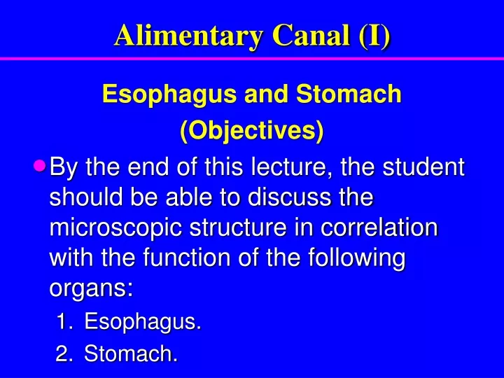
Alimentary Canal (I) Esophagus and Stomach (Objectives) • By the end of this lecture, the student should be able to discuss the microscopic structure in correlation with the function of the following organs: • Esophagus. • Stomach.
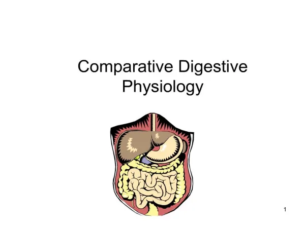
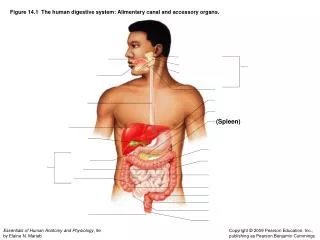
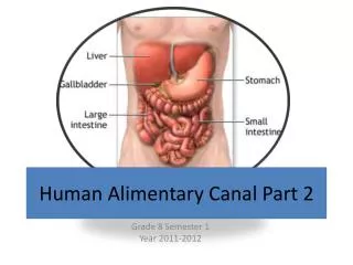
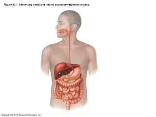
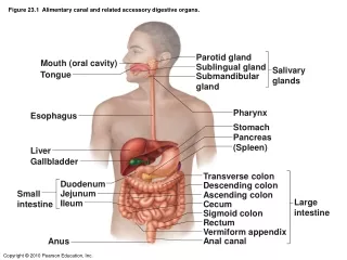
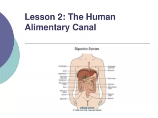
Alimentary Canal • Is the tubular portion of digestive system. • Is subdivided into: esophagus, stomach, small intestine (duodenum, jejunum and ileum), and large intestine (cecum, colon, rectum, anal canal, and appendix).
General Architectureof L/M Structure of Alimentary Canal 1- Mucosa. 2- Submucosa. 3- Muscularisexterna. 4- Adventitia OR serosa. Serosa
General Architectureof L/M Structure of Alimentary Canal or Serosa
Esophagus Four concentric layers: • Mucosa: • Epithelial Lining:Non-Keratinized Stratified Squamous Epithelium. • Lamina propria:Loose areolar C.T. with mucosal esophageal glands (secretion of mucus) in the upper and lower ends. • Muscularismucosae:Few layers of smooth muscle fibers. Serosa
Esophagus • Submucosa: • Loose areolar C.T. containing blood vessels, nerves, submucosal esophageal glands(secretion of mucus)& • Meissner’s plexus of nerve fibers and nerve cells. • MuscularisExterna: Two muscle layers: • Inner circular layer. • Outer longitudinal layer. • Upper 1/3: both layers are skeletal M. • Middle 1/3: inner layer is smooth muscle outer layer is skeletal M. • Lower 1/3: both layers are smooth M. • Auerbach’s (myenteric) plexus in between the 2 layers
Esophagus • Serosa or Adventitia: • Adventitia: is loose areolar C.T. not covered by mesothelium. • Serosa: is loose areolar C.T. covered by mesothelium (simple squamous epithelium) in the abdominal part of the esophagus. Serosa
STOMACH • It has 4 regions: cardia, fundus, body and pylorus. • Mucosa has folds, known as rugae that disappear in the distended stomach. fundus cardia body pylorus
Fundus (and Body) of Stomach • Mucosa:is invaded by fundic glands. The surface epithelium of the mucosa is simple columnar mucus-secreting cells. • Submucosa: • Connective tissue containing blood vessels, nerves, and Meissner’s plexus. • NO glands. • MuscularisExterna: • Three smooth muscle layers: • Inner oblique. • Middle circular. • Outer longitudinal. • Auerbach’s(myenteric) plexus. • Serosa: • C.T. covered by mesothelium.
Mucosa of Fundus of Stomach • It is composed of: 1. Surface Columnar Epithelium: Simple columnar epithelium: secretes mucus. 2. Lamina propria: C.T. invaded by numerous fundic glands with lymphoid elements. 3. Muscularis mucosae: 2 layers of smooth muscle fibers.
Mucosa of Fundus of Stomach Surface Columnar Epithelium
Fundic Glands • Fundic glands have: • Short pits: one fourth of mucosa. • Simple branched tubular glands. • Are rich in parietal & chief cells.
Mucosa of Fundus of Stomach 1 2 Lumen. Surface columnar epithelium. Pits of fundic glands. Fundic glands. Lamina propria. Muscularis mucosae. 3 4 5 6
Fundic Glands Composed of 5 cell types: • Parietal (oxyntic) cells. • Peptic (chief) cells. • Mucous neck cells. • Enteroendocrine(EE, DNES) cells. • Stem cells.
Fundic Glands • Parietal (oxyntic) cells: • Shape: pyramidal or polygonal. • Nucleus: central, round. • Cytoplasm: • deeply acidophilic, rich in SER and mitochondria (40% of the cell volume). • C-shaped intracellular canaliculus. • Secrete HCl and gastric intrinsic factor that helps absorption of vitamin B12. • Parietal - why? • Oxyntic - why?
Fundic Glands 2. Peptic (chief) cells: • The predominant cell type. • Columnar cells. • Nucleus: basal, round. • Cytoplasm: • basohilic with apical secretory granules. • secrete pepsinogen.
Fundic Glands 3. Mucous neck cells:secrete mucus. 4.Enteroendocrine (EE) (DNES) cells:secrete hormones (e.g. serotonin, endorphin). 5. Stem cells: regenerative cells.
Pylorus of Stomach • Mucosa: is invaded by pyloric glands. The surface epithelium is simple columnar mucus-secreting cells. • Submucosa: • Connective tissue containing blood vessels, nerves, and Meissner’s plexus. • NO glands. • MuscularisExterna: • Two smooth muscle layers: • Inner circular. • Outer longitudinal. • Auerbach’s plexus. • Serosa: • C.T. covered by mesothelium Lumen Surface epithelium Pits of pyloric glands Lamina propria Muscularis mucosae Submucosa Muscularis externa
Pyloric glands • Their pits are deep --- about half the length of mucosa. • They are branched and convoluted --- many cross sections.
Pyloric glands • Cells of pyloric glands: • Mucus neck cells (Mucus secreting cells): • The predominant cells. • Secrete mucus. • EE cells: • EC cells • G cells • D cells • A cells • Stem cells. • Parietal cells: few. • No peptic cells.