Download
1 / 35
350 likes | 370 Views
Respirastory system. D. L. Kiss Anna Semmelweis University Department of Anatomy , Histology and Embryology 2018. Extrapulmonar : Nasal cavity - nasopharynx Larynx Trachea Intrapulmonar : Bronchi : principal lobar segmental Bronchioles : terminals respiratory

E N D
Respirastorysystem D. L. Kiss Anna Semmelweis University Department of Anatomy, Histology and Embryology 2018.
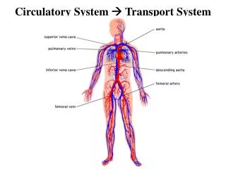
Extrapulmonar: • Nasalcavity - nasopharynx • Larynx • Trachea • Intrapulmonar: • Bronchi: principal lobar segmental • Bronchioles: terminals respiratory • Repiratory part: alveoli
Nasalcavity Olfactoryarea: receptors Vestibulum: skin Regiorespiratorica: pseudostratifiedciliated epithelium (gobletcells)
Regiorespirtorica: Propria: seros+mucus glands vestibulum
cilia cilia Olfactoryregion Receptors: primary sensory cells: bipolar cells • contain OBP (odorant binding protein) receptors on their plasma membrane • axon-like processes - fila olfactoria – bulbus olfactorius • the number of them: 2 x 10⁷ • life time: 30-60 days • more than 100 receptor molecules bulb-like ending supporting cell receptor basal cells basement membrane Schwann cells axon
nasopharynx tongue oropharynx softpalate laryngopharynx epiglottis
Waldeyer’slymphatic ring Pharynx: parsnasalis: • tonsillapharyngea+tonsillaetubariae(pseudostratifiedciliatedepithelium) p. oralis: • tonsillapalatina (stratified non- keratinizedepithelium) • tonsillaelinguales (stratified non- keratinizedepithelium)
Larynx epiglottis aryepiglottic fold vestibular fold vestibulum ventriculus infraglotticcavity vocal fold (vocalcord)
Trachea anteriorview posteriorview left right left Right Principalbronchi
Bronchustree Principalbronchuslobalbronchus segmentalbronchusterminalbronchus bronchiolusterminalbronchiolus bronchiolusrespiratoricussacculus: ductus alveolaris alveoli
Histologicalchangesintherespiratorysystem • cartilagedisappears: gradually • glandsaredisappearing • smoothmusclebecomescontinuous (broncioles) and thendisappears • epitheliumbecomesthin
Branches of theterminalbronchi, sacculi and alveoli Lobes : segments Segments: borders: veins artery + bronchioles (centrally)
Alveoli and alveolarsepti macrophage alveolarentrance elasticfibers type I cells type II cells macrophageintheseptum capillary
Pneumocytes: simplesquamousepithelialcells Type I cells: flat, squamouscells: gassexchange Type II cells: surfactantsecretion: decreasesthesurfacetension
Surfactant Decreasesthealveolarsurfacetension, activelyparticipate in theclearance of foreignmaterials Lowsurfacetension: increasestheflexibility of thelung makesthealveoli stabil e Absence of thesurfactant: alveolicollapse
Respiratorydistresssyndrome (RDS) – functionalsurfactantdisorder • Fetalorneonatal RDS (IRDS): more frequent in prematureinfant 1.) unmaturedbiosyntheticpathways 2.) inactivation of thesurfactant (intraalveolarcoagulation) 3.) increaseduse of surfactantcausedbychronicrespiratorymechanism 4.) injury of thebiosyntheticpathway (acidosis, decreasedpulmonaryblood flow) • Adult RDS (ARDS):acutrespiratoryinsufficiency activation of neutrophylgranulocytes and alveolarmacrophagesrelease of permeabilityincreasingsubstances (shock, trauma:. burning, infections, inhalation of toxicgases, overdosage of drogssetc)
Development of the lung • 4 weeksembryo: respiratorydiverticulum:outgrowth of theventralwall of theforegut; • in themesodermretinoidacidproduction is increasing • in theendoderm TBX4 transcriptionfactorproduction • TBX4: inducesappearance, increase and differentiation of thelung primordia • epithelium: endoderm • connectivetissue, cartilage and muscle: mesoderm
Development of thelung 1.) respiratorydiverticulum communicateswiththeforegut 2.) Tracheoesophagealridge: dorsal: esophagus ventral: trachea 3.) 2 bronchusbuds atthe 5th week: - right and leftprincipalbronchi - then: right: 3 lobarbrobnchi, left: 2 lobarbronchi - dichotomicdivision: segmentalbronchi
Bibliography • Snell RS, ClinicalAnatomy, Little, Brown & Co, Boston, 1995 • Moore KL, Dalley AF: Clinically Oriented Anatomy, Lippincott, 1999 • Sobotta: Atlas of Human Anatomy • Röhlich: Szövettan