Download
1 / 24
430 likes | 1.38k Views
Chlamydia. Prof.Dr.Rıza Durmaz YBÜ-2014. 1) Genus : Chlamydia Chlamydia trachomatis. 2) Genus : chlamydophila C.pneumoniae C.psittaci. Classification of the family Chlamydiaceae. The family Chlamydiaceae consists of two medically important genera.

E N D
Chlamydia Prof.Dr.Rıza Durmaz YBÜ-2014
1)Genus: Chlamydia Chlamydiatrachomatis 2) Genus: chlamydophila C.pneumoniae C.psittaci Classification of thefamilyChlamydiaceae The family Chlamydiaceae consists of two medically important genera • Chlamydiae are obligate intracellular parasites • The members of this group share a unique development cycle, a common morphology and a common family antigen. • They are not transmitted by arthropods
Chlamydiaceae • Theyaresmallranging in size from about 0.2µmto lµm. • They can pass 0.45 micrometerfilters • Theyareobligateintracellularparasites • theywereconsideredviruses • Theyhavethefollowingproperties of bacteria • Possessinnerandoutermembranessimilarto Gram (-) bacteria • Containboth DNA and RNA • Theyhaveprokaryoticribosomes • Theysynthesizetheirownproteins, nucleicacidsandlipids • Theysusceptibletonumerousantibacterialantibiotics. • Theylack a typicalbacterialpeptidoglycanlayer in cellwall
Physiology and structure • Chlamydiaehavegenus-specificlipopolysaccaride (LPS) antigen in theircellwall. • Theirmajoroutermembraneproteins (MOMP) arespecies- andstrain- specific • Theyareenergyparasites, usinghost ATP • Theygrow in cultures of a variety of eukaryoticcellslines • McCoycells, HL, or Hep-2 cells. Alltypes of chlamydiaeproliferate in embryonatedeggs.
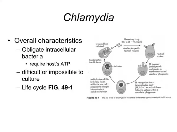
AllChlamydiaehave a commonreproductivecycle, forming • Elementary body; • Infectious form, metabolically inert • Extracellular spore-like state • Reticulate body; • Non-infectious form, metabolically active • obligate intracellular form in eukaryotic cells • 48-72 hour cycle
Growthcycle • Thegrowthcycleinitiatewhenthesmall (300-400nm) EBsattachedtothemicrovilli of susceptiblecells, • stimulateactivepenetrationintothehostcell. • Afterinternalization, chlamydiaremain in cytoplasmicphagosomes, • Fusion of cellularlysosomeswith EB-containingphagosomeandintracellularkillingis inhibited • In 6-8 hours, theEBsreorganiseintothelarger (800-1000 nm)metabolicallyactiveRBs • RBsreplicatebybinaryfission (continuous 18-24 h)
Growth cycle-2 • Histologicstains can detectthephagosomewithaccumulatedRBs, calledinclusion. • 18-24 h afterinfection, RBsbeginreorganizingintothesmallerEBs • Between 48-72h, hostcellrupturesandthenreleasetheinfectiveEBs.
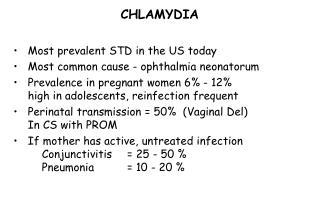
Chlamydia • Theymaycolonizeandinfecttissues of theeyeandurogenitaltractin humans. • Chlamydiatrachomatiscausesseveralimportantdiseases in humans: • lymphogranulomavenereumsexuallytransmitteddisease • trachoma, a leadingcause of blindnessworldwide • Chlamydiapneumoniae • a cause of pneumonia • has beenrecentlylinkedtoatherosclerosis.
Rickettsiaceae Family • Includes the genera: • RickettsiaandOrientia • Obligate intracellular Gram negative bacteria
Rickettsia and Orientia • Theyweresmall,( 0.3 x 1-2 µm) • Stainedpoorlywith Gram stain; bestwithGiemsaorGimenez • Theygrowonly in eukaryoticcells (intracellularparasites)
Characteristics • Cell wallstructuresaresimilarto Gram (-) rods • Peptidoglycanlayer is minimal • LPS has weakendotoxinactivity. • Orientialacksbothpeptidoglycanlayerand LPS • Rickettcia is surroundedwithlooselyadherentslimelayer • Theycontain DNA, RNA andenzymesforKrep’scycleandribosomesfor protein synthesis • Multiplicationbybinaryfission • Theyareinhibitedbyantibiotics (e.g.tetracyline, chloramphenicole)
7. PathogenicspeciesRickettsiaandOrientiaaremaintainedin animalandarthropodreservoirs • Theyaretransmittedbyarthropodvectors(e.g.ticks,mites, liceandfleas) • Humansareaccidentalhosts • Theyproducediseasessuch as typhusfever, RockyMountainSpotted Fever,
Growthcycle • The bacteria enter eukaryotic cells by attaching to host cell surface receptors and stimulating phagocytosis. • After engulfment, Rickettsia and Orientiadegrade the phagosome membrane by producing a phospholipase and must be released into the cytoplasm, or the organism will not survive. • Multiplication in the host cell by binary fission is slow (generation time, 9 to 12 hours). • Orientia and the spotted fever group of Rickettsia grow in the cytoplasm and nucleus of infected cells and are continually released from cells • In contrast, the typhus group accumulates in the cell cytoplasm until the cell membranes lyse, bacterial release. • Once these bacteria are released from the host cell, they are unstable and die quickly.
Ehrlichia, AnaplasmaandCoxiella • Members of the families Anaplasmataceae and Coxiellaceae are intracellular pathogens.
Characteristics of Ehrlichia, Anaplasma • Theyareintracellularbacteria • Theyparasitizemononuclearorgranulocyticphagocytes, erithrocytesandplatelets • Infection of hematopoieticcells • Thecellwallstructure of EhrlichiaandAnaplasma is similartothat of Gram (-) bacteria, but peptidoglycanand LP are not present. • Theysurvivewithin a cytoplasmicvacuolein theinfectedarthropodormammaliancell.
Growthcycle • After entry into the host cell, theyremain in the phagocytic vacuole • Fusion with lysosomes is prevented • The bacteria can multiple by binary fission in the phagosomewithout exposure to the hydrolytic lysosome enzymes. • Two morphologic forms of the bacteria exist: • Small (0.2 to 0.4 µm) elementary bodies and • Larger (0.8 to 1.5 µm) reticulate bodies. • A few days after the cell is infected, the replicating elementary bodies assemble into membrane-enclosed masses called morulae. • Detection of morulae when the cells are stained with Giemsa or Wright stains is a rapid, specific diagnostic test Multiple morulae of Ehrlichia canis in DH82 tissue culture cells
Coxiella • Coxiellaburnetii is classified in Coxiellaceae • It has gram-negative cellwall, • Itstains weakly with the Gram stain, • Itgrow intracellularly in eukaryotic cells • Two structural forms of C. burnetii are recognized: • small cell variants that are resistant to environmental stress (e.g., heat, desiccation, chemical agents) and large cell variants that are the metabolically active form. • Human infections occur after the inhalation of airborne particles from a contaminated environmental source or, less commonly, after ingestion of contaminated unpasteurized milk or other dairy products. • Ticks do not transmit disease to humans..
Mycoplasma and Ureaplasma • Three human pathogens • Mycoplasma pneumoniae • M. hominis • Ureaplasma urealyticum
Physiology and structure • Thesmallestfree-livingbacteria (0.1-0.3 µm). • Theydonthave a cellwall, • Theircellmembranecontainssterols. • Mycoplasmaresistanttopenicillins, cephalosporins, vancomycinandotherantibiotics (thecellwallinhibitors) • Theymay be free-living in soilandsewage, • Theymay be inhabitants of themouthandurinarytract of humans, orpathogens. • Inhumans, Mycoplasmapneumoniaecausesprimaryatypicalpneumonia, alsocalledwalkingpneumonia.
Physiology and structure • Mycoplasmas form pleomorphicfilaments • They can passthroughthe 0.45-µm filtersusedtoremovebacteriafromsolutions • Organismsdividebybinaryfission • Theygrow on artificialcell-freemedia • Theycontainboth RNA an DNA • Theyarefacultativeanaerobic (exceptM.pneumoniaewhich is strictaerobes) • Theyrequireexogenoussterolssuppliedbyanimal serum addedtothegrowthmedium. • Theygrowslowly, gen.time 1-6 hours • They form smallcoloniesthathavea fried –eggappearance
Physiology and structure • M.pneumoniae is an exception, itscolonieshavebeendescribed as mulberryshaped • Colonies of Ureoplasmaareextremelysmallmeasuring 10-50µm • majorantigenicdeterminantsaremembraneglycolipidsandproteins. Theseantigenscross-reactwithhumantissuesandotherbacteria.