Download
1 / 33
330 likes | 477 Views
Skin and the Integumentary System. Integumentary System. Cutaneous membrane and certain accessory organs Epithelium and connective tissue. Types of Membranes. Serous: line body cavities that lack outside openings Thorax, abdomen Simple squamous epithelium and loose connective tissue

E N D
Integumentary System • Cutaneous membrane and certain accessory organs • Epithelium and connective tissue
Types of Membranes • Serous: line body cavities that lack outside openings • Thorax, abdomen • Simple squamous epithelium and loose connective tissue • Secrete watery serous fluid: lubricated membrane surface
Types of Membranes • Mucous : line cavities and tubes that open to the outside • Oral and nasal cavities • Tubes of digestive, respiratory, urinary, and reproductive systems • Epithelium overlying loose connective tissue • Secrete mucus
Types of Membranes • Synovial: form inner lining of the joint cavities • Between bones and joints • Dense connective tissue over loose connective tissue and adipose tissue • Secrete a thick, colorless synovial fluid into joint cavity • Lubricated the ends of bones within joint
Types of Membranes • Cutaneous membrane: skin • One of the larger and more versatile organs • Vital for homeostasis • Functions • Protective covering • Regulate body temperature • Retards water loss from deeper tissue • Houses sensory receptors • Synthesizes various biochemicals • Excretes small quantities of waste
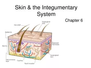
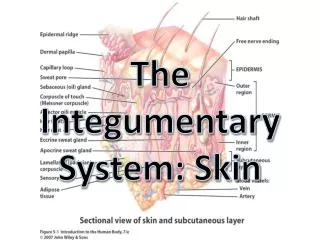
Skin • Epidermis: outer layer • Stratifies squamous epithelium • Dermis: inner layer • Thicker • Connective tissue: collagenous and elastic fibers • Epithelial, smooth muscle, & nervous tissue • Blood • Basement membrane • Subcutaneous Layer: hypodermis • Masses of loose connective tissue and adipose tissue • Bind skin to underlying organs
Epidermis • Lacks blood vessels • Stratum basale: deepest layer • Nourished by dermal blood vessels • Cells divide and grow • older epidermal cells are pushed toward the surface • Further to the surface fewer nutrients • eventually they die
Skin • Kertinocytes: older cells • Keratinization: process of hardening the cells • Cytoplasm fills with keratin protein • Tough, waterproof fibers • Stratum corneum: layers of tough, dead skin on the surface
Epidermis • Functions • Shields against excessive water lose • Mechanical injury • Effects of harmful chemicals • Protection against disease causing agents • Melanocytes: cells that produce melanin • Skin color pigment • Absorbs light energy and lessens effects of UV radiation • Lie in deepest part of epidermis and dermis tissue • Cytocrine secretion: transport of melanin to epidermal cells
Skin Color • Due to melanin • All people have about the same number of melanocytes • Genetic • Variations in color • Amount of melanin produce • Distribution and size of pigment granules • Sunlight, UV light, X-rays stimulate production of extra melanin • Oxygen Content of blood • Highly oxygenated bright red blood appear pinkish • Less oxygenated dark red blood appear bluish • cyanosis
Dermis • Binds the epidermis to underlying tissues • Network of these fibers give the skin toughness and elasticity • Dermal blood vessels supply nutrients for all skin cells • Regulate body temperature
Dermis • Nerve Fibers • Motor Fibers: impulses from brain or spinal cord to dermal muscles and glands • Sensory fibers: carry impulses away from specialized sensory receptors • Touch receptors • Hair follicles, sebaceous glands, and sweat glands
Subcutaneous Layer • Hypodermis: beneath the dermis • Loose connective tissue • Adipose tissues • Insulated heat • Contains major blood vessels
Hair Follicles • Present on all skin surfaces except the palm, soles, lips, nipples, and parts of external reproductive organs • Hair Follicle: tubelike depression from which the hair develops • Extends to the dermis and contains the hair root • Nourished from dermal blood vessels • With division and growth, older cells are pushed toward the top • Become keratinized as they move up • Shaft: structure that extends from skin surface • Dead epidermal cells
Hair Follicles • Arrector pili muscle: smooth muscles attached to each hair follicle • Short hair stands on end when muscles contract • Emotional upheaval or cold • Goose bumps • Hair color: genetic • Amount of pigment the epidermal melanocytes produced • Dark: a lot of pigment • Blond: intermediate quantity • White: no pigment • Red: trichosiderin
Sebaceous Glands • Groups of specialized epithelial cells • Usually associated with hair follicles • Holocrine glands that secrete fluid through small ducts in hair follicles • Sebum: secrete an oily mixture of fatty material and cellular debris • Skin and hair are kept soft, pliable, and waterproof
Nails • Protective coverings on the ends of fingers and toes • Keratinized stratified squamous epithelial cells • Nail Root: where nail originates • Near the nail’s proximal end • Lunula: whitish, half-moon shaped area • Most active growing region • Nail Bed: epithelium that nail slides over as it grows • Remains attached
Sweat Glands • Sudoriferous Glands • Widespread exocrine glands • Deeper dermis or superficial subcutaneous layer • Tiny tube that originates as a ball-shaped coil • Coiled portion of the gland is closed and is lined with sweat-secreting epithelial cells
Types of Sweat Glands • Eccrine Glands: most numerous • Respond to temperature • Forehead, neck, and back • Sweat is carried to a pore • Mostly water with small quantities of salts and wastes • Apocrine Glands: activate with emotional distress, fright, or pain • Axillary regions and groin • Connect to hair follicles • Ceruminous Glands: ear wax • Female mammary glands: milk
Regulation of Body Temperature • Slight shifts in body temperature disrupts metabolic reaction rates • Deep body temp.: 37ºC or 98.6ºF • Maintain by balancing heat lose with heat gain • Heat: product of cellular metabolism • Higher activity more heat produced • Skeletal and cardiac muscle and liver cells
Intense Heat • Nerve impulses stimulate structures in the skin and other organs to release heat • Physical exercise: muscle release heat to the blood which is carried away • Blood vessels dilate and heat escapes to outside • Eccrine Sweat glands become active • Sweat evaporates cools the surface
Cold • If too much heat is lost: • Muscles in the walls of dermal blood vessels are stimulated to contract • Decreases blood flow through the skin • Sweat gland remain inactive • Skeletal muscle fibers contract • Increase rate of cellular respiration heat is a byproduct • Small groups of muscles may contract rhythmically with more force, shiver • Generates more heat
Wound Healing • Inflammation: area around a wound becomes red and painfully swollen • Blood vessels dilate and become more permeable • Forces fluids to leave the blood vessels and enter damaged tissue • Provides more nutrients and oxygen to aid healing • Extent of healing depends on extent of injury
How We Heal • Shallow Skin Break: Epithelial cells along the margin are stimulated to divide more rapidly • Newly formed cells fill the gap
Deep Cut • Injury to dermis or subcutaneous layer • Blood vessels break and escaping blood forms a clot • Scab: blood clot and dried tissue fluids • Covers and protects underlying tissue • Fibroblast migrate into region and form new collagenous fibers that bind wound edges • Closing large wounds speeds the process
Continuing to Heal • Blood vessels extend to area beneath the scab • cells remove dead cells and other debris • Damaged tissues are replaced • Scab sloughs off • Scar: with extensive wounds, newly form tissue different than surroundings
Large Wounds • Granulations: formation of small, rounded masses in the exposed tissue • New branch of a blood vessel • Cluster of collagen-secreting fibroblasts • In time, fibroblasts migrate away and blood vessels are reabsorbed • Leaves a scar