Download
1 / 103
1.16k likes | 1.75k Views
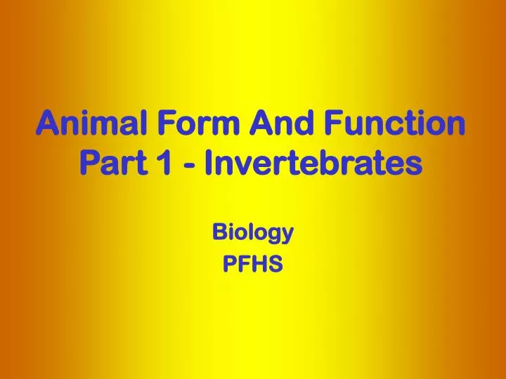
Animal Form And Function Part 1 - Invertebrates. Biology PFHS. Lecture Outline: What is an animal? Origin of animal body form diversity Correlation between form and function Organization of body plans into grades Views of animal diversity. What is an animal? How do we define an animal?

E N D
Animal Form And FunctionPart 1 - Invertebrates Biology PFHS
Lecture Outline: What is an animal? Origin of animal body form diversity Correlation between form and function Organization of body plans into grades Views of animal diversity
What is an animal? How do we define an animal? While there are exceptions to nearly every criterion for distinguishing an animal from other life forms, the following criteria, when taken together, create a reasonable definition.
1. Animals are multicellular eukaryotes. 2. Animals are Heterotrophs: Cannot make their own food - cannot convert simple inorganic molecules into complex organic molecules. They must take in preformed organic molecules through ingestion, eating other organisms or organic material that is decomposing. Plants are Autotrophs – they can synthesize their nutrition from simple inorganicmolecules into complex organic molecules.
3. Animal cells lack cell walls that provide structural supports for plants and fungi. Cell Membrane: Every cell is enclosed in a membrane. The membrane is a double layer of lipids (lipid bilayer) but is made quite complex by the presence of numerous proteins that are important to cell activity. Cell Wall: Prokaryotic cells, fungi cells, and plant cells have a rigid cell wall in addition to the cell membrane that is made up of polysaccharides. The cell wall provides and maintains the shape of these cells and serves an extra protective barrier. Animal Cell Plant Cell
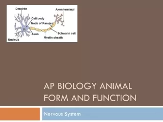
4.Locomotion: Animals are capable of moving from one place to another. This complex function is achieved by coordinated functioning of two unique tissue types. i. Nervous tissue for impulse conduction ii. Muscle tissue for movement. Exception: Some animals do not have locomotor ability – sedentary.
5. Reproduction: Most animals reproduce sexually, with the diploid stage usually dominating the life cycle. In most species, a flagellated haploid sperm fertilizes a haploid egg. The fertilized diploid egg - zygote - undergoes cleavage, a succession mitotic cell divisions, leading to the formation of a multicellular, hollow ball of cells called the blastula.
6. Characteristics that are true for most animals, but not all… • The multicellular bodies of animals are held together with the extracellular proteins, especially collagen. • In addition, other structural proteins create several types of intercellular junctions, including tight junctions, desmosomes, and gap junctions, that hold tissues together. • Animals have skeleton – endoskeleton (bones) or exoskeleton (shells) – that provides rigidity of form.
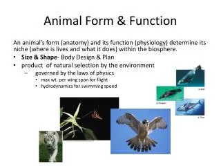
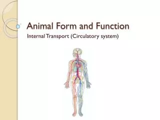
Correlation between Form and function Animals show a correlation between body form (structure) and function. Form fits function at all the levels of life, from molecules to organisms. Knowledge of a structure provides insight into what it does and how its works. Conversely, knowing the function of a structure provides insight about its construction. Therefore, sometimes distinction between anatomy and physiology is blurred.
Animal Classification: • Living animals are classified in to 35 different phyla based body plans. • Most are marine phyla, only a few terrestrial phyla – most species diversity is in the ocean. We will cover about 13 of these phyla • Four basic features of animal body plan are the basis for classification of animals into various phyla.: Body symmetry Germ Layer • Coelom Development pattern
Body Symmetry: • Mainly based on anatomical features in adults and certain details of embryonic development. • The symmetry of an animal generally fits its lifestyle. • i. Radial Symmetry • ii. Bilateral Symmetry • iii. Asymmetry (not common)
i.Radial Symmetry: • Many radial animals are sessile or planktonic and need to meet the environment equally well from all sides. Multiple planes of symmetry
ii. Bilateral symmetry: • Animals that move actively are bilateral. • Head, anterior end, encounters the environment -food, danger, and other stimuli. • Bilateral symmetry is associated with cephalization, concentration of sensory equipment on the anterior end – head. • Cephalization leads to the development of central nervous system. Dorsal Posterior Single plane of symmetry Anterior Ventral
2. Germ Layers: Three germ layers: Ectoderm: forms the outer covering and, the central nervous system (if present). Endoderm: lines the digestive tract and the organs derived from it, such as the liver and lungs of vertebrates. Mesoderm: lies between the endoderm and ectoderm and develops into muscles and other organs e.g., kidneys and gonads. i. Diploblastic:have two germ layers – ectoderm and endoderm. e. g., the radiata. ii. Triploblastic: have three germ layers – ectoderm, endoderm and mesoderm. e. g., the bilateria
3. Coelom: The germ layers form a cavity around the internal organs – Coelom. • Coelom has many functions. • Its fluid cushions the internal organs, helping to prevent internal injury. • The noncompressible fluid of the body cavity can function as a hydrostatic skeleton against which muscles can work. • The present of the cavity enables the internal organs to grow and move independently of the outer body wall.
A coelom – the body cavity - could be a true coelom or a false coelom. • Based on the type of coelom and presence or absence of a coelom and animal could be classified as: • Coelomate • Pseudocoelomate (false coelom) • Acoelomate (no coelom)
Coelomates are organisms with a true coelom, a fluid-filled body cavity completely lined by mesoderm. • The inner and outer layers of tissue that surround the cavity connect dorsally and ventrally to form mesenteries, which suspend the internal organs.
Pseudocoelomates, have a body cavity, but it is not completely lined by mesoderm, e.g., rotifers (phylum Rotifera) and the roundworms (phylum Nematoda).
Acoelomates have a solid body and lack a body cavity, e.g., (the phylum Platyhelminthes) .
4. Development pattern: DEUTEROSTOMES PROTOSTOMES (a) Cleavage (zygote divides into ball of cells) Spiral cleavage Radial cleavage (b) Gastrulation (cells invaginate to form gut) Mouth Pore becomes mouth Pore becomes anus Anus (c) Coelom formation Gut Gut Coelum Mesoderm Mesoderm Block of solid mesoderm splits to form coelom Mesoderm pockets pinch off of gut to form coelom
Major Steps in Animal Form and Function Choanoflagellates Poriferans Protist ancestor Ectoderm Multi-cellularity Endoderm Cnidarians Ectoderm Mesoderm Endoderm Two tissue layers Protostomes Anus Developing mouth Ectoderm Mouth Body cavity Mesoderm Endoderm Deutero- stomes Three tissue layers, a body cavity, and bilateral symmetry Anus New forms of embryological, development, including the formation of the mouth secondarily
Porifera • Poriferans are commonly referred to as sponges. • fossil sponges are among the oldest known animal fossils, dating from thelate precambrian. • The approximately 5,000 living sponge species are classified in the phylum Porifera
Porifera • Sponges are characterized by the possession of a feeding system unique among animals. • Poriferans don't have mouths; instead, they have tiny pores in their outer walls through which water is drawn. • Cells in the sponge walls filter goodies from the water as the water is • pumped through the body and out other larger openings. The flow of • water through the sponge is unidirectional, driven by the beating of • flagella which line the surface of chambers connected by a series of • canals. • Sponge cells perform a variety of bodily functions and appear to be • more independent of each other than are the cells of other animals.
(b) Central cavity Gelatinous middle layer with skeletal materials and motile cells that can become gametes Motile cell Microvilli Collar cell Pore Central cavity Skeletal material (spicule) Collar cells are similar in form to choanoflagellate protists, suggesting that animals derived from colonial choanoflagellates Fig. 13.04
Porifera • Form symbiotic relationships with bacteria and fungi • Produce a wide array of chemical defense compounds • Useful for Medicinal lead compounds • Discodermalide a potent anticancer compound in clinical trials
Introduction to Cnidaria • Jellyfish, corals, and other stingers. . . • Cnidarians are incredibly diverse in form, as evidenced by colonial siphonorphores, massive medusae and corals, feathery hydroids, andbox jellies with complex eyes. • Yet, these diverse animals are all armed with stinging cells called nematocysts. Cnidarians are united based on the presumption that their nematocysts have been inherited from a single common ancestor. • There are four major groups of cnidarians: • Anthazoa - which includes true corals, anemones, and sea pens • Cubozoa - the amazing box jellies with complex eyes and potent toxins • Hydrozoa - the most diverse group with siphonophores, hydroids, fire corals, and many medusae • Scyphozoa - the true jellyfish.
CnidariaThere are four major groups of cnidarians: • Anthazoa - which includes true corals, anemones, and sea pens
Coral Reef Ecosystems • Coral reefs and other marine ecosystems, however, contain more varied life forms than do land habitats. • All but one of the world's 35 phyla are found in marine environments -- 15 exclusively so. • Coral reefs are found in shallow waters, extending to depths of 30 meters and cover 15 percent of the world's coastline (0.2% of the total area of oceans) • Fish production on these reefs and on the adjacent continental shelf could amount to nearly 10 percent of global fisheries production if fully exploited. • Coral reefs also protect coastal areas from erosion. In the case of coral atolls, coral provides the foundation of the island itself. In the Indian Ocean, 77 percent of isolated islands and island archipelagoes are built exclusively of reef depositions.
Affiliated Ecosystems • Coral reefs stand out from other marine environments because of their species diversity, but many coral reef species also depend on other affiliated ecosystems. • Often, coral reefs, mangroves, and sea grass beds are linked physically and biologically: • reefs serve as breakwaters that allow coastal mangroves to develop; • the calcium of the reef provides the sand and sediment in which mangroves and sea grasses grow; and • the mangroves and sea grass communities provide energy input into the coastal ecosystem and serve as spawning, rearing, and foraging habitat for the many of the species associated with the reefs.
Coral Reefs Mangroves Sea Grass Beds
Coral Reef Ecosystems Some Facts and Figures on Coral Reefs as a Fishery Resource • Healthy coral reefs can produce more than 20 metric tons of fish and other edible marine products per square kilometer per year. Unfortunately, 95% of the country's coral reefs are various stages of deterioration due to destructive and illegal practices, siltation and sedimentation, and pollutants from industrial and domestic wastes and construction activities, among others. The Destruction of Coral Reefs • Fishing practices destructive to coral reefs include muro-ami, cyanide fishing, trawl fishing, indiscriminate harvesting, and dynamite fishing. Blast or dynamite fishing alone destroys an average of one to three square meters of corals per blast. consider this -- coral grows very slowly at an average of one to two inches a year. Thus, it takes hundreds of years to grow a coral reef!
CnidariaThere are four major groups of cnidarians: • Hydrozoa - the most diverse group with siphonophores, hydroids, fire corals, and many medusae
CnidariaThere are four major groups of cnidarians: • Scyphozoa – the true jellyfish
CnidariaThere are four major groups of cnidarians: Cubozoa - the amazing box jellies with complex eyes and potent toxins
(e) Radial body plan Jellyfish, from top Nerve cell Nematocyst discharged (f) Tentacle Nematocyst- bearing cell Mouth Epidermis Epidermis Gastrodermis Mesoglea Mesoglea Mouth Digestive enzyme- secreting cell Gastrovascular cavity Muscle cells Fig. 13.05 Slide 4 Polyp form Medusa form
Treatment for Stings • Primary first aid for any jellyfish sting should be to minimize the number of nematocysts discharging into the skin and to reduce the harmful effects of the venom. If stung by a jellyfish, the victim should carefully remove the tentacles that adhere to the skin by using sand, clothing, towels, seaweed or other available materials. As long as tentacles remain on the skin, they will continue to discharge venom. • A variety of substances have been used to reduce the effects of jellyfish stings. Meat tenderizer, sugar, vinegar, plant juices and sodium bicarbonate have all been used with varying degrees of success. Methylated spirits and other forms of alcohol formerly recommended for inhibiting stinging cells actually stimulate them and may increase pain and cause severe skin reactions. Picric acid and human urine also cause a discharge of nematocysts and should not be used. Victims of serious stings should make every effort to get out of the water as soon as possible to avoid drowning. If swelling and pain from more serious stings persists, prompt medical attention should be sought. Recovery periods can vary from several minutes to several weeks.
Adult female medusa Tentacle Gonad Adult male medusa Egg Sexual reproduction Zygote Sperm Development New medusa Planula larva Cilia Asexual reproduction Stack of developing medusae Differentiation Hydra-like polyp Fig.13.06
Protostomes Fruit fly larva, cross section First opening becomes the mouth Anus Fig. 13.08a
Platyhelminthes • Reproduction is by both sexual and asexual means. For sexual reproduction, planarians are hermaphroditic organisms. • Asexual reproduction is accomplished by fragmentation followed by regeneration, and many of these free-living flatworms have remarkable regenerative abilities. As a result of this, the turbellarian flatworms have contributed a great deal of information to our knowledge of regeneration.
Hermaphrodite • An organism which has both male and female organs, and produces both male gametes (sperm) and female gametes (eggs). • The organism can have both types of organs at the same time (simultaneous hermaphrodite) or have one type early in life and the other type later in life (sequential hermaphrodite).
Platyhelminthes Class Trematoda (Flukes) • The flukes are all parasitic organisms; the adult stage living in or on a wide variety of vertebrate animals. Of the groups the blood flukes are the most important as they are the causative agents for the human disease known as Schistosomiasis. This debilitating disease affects some 400,000,000 persons in Asia, Africa, parts of South America and the Caribbean Class Cestoda (Tapeworm) • Tapeworms are elongate, intestinal parasites consisting of an anterior scolex for attachment and numerous body segments called proglottids. They are highly modified to a parasitic existence and have no digestive system. Tapeworms also have complicated life histories.
Tape Worms • The adult lives in the small intestine. It is hooked onto the intestinal wall by a structure called a rostellum which is sort of like a hat with hooks on it. The tapeworm also has six rows of teeth to grab on with. • Once docked like a boat to the host intestinal wall, the tapeworm begins to grow a long tail. • The tapeworm absorbs nutrients through its skin as the food being digested by the host flows past it. • Older segments are pushed toward the tip of the tail as new segments are produced by the neckpiece. By the time a segment has reached the end of the tail, only the reproductive tract is left. When the segment drops off, it is basically just a sac of tapeworm eggs. 28 foot tape worm
(b) The flatworm body plan Dorsal Posterior Left Head Right Anterior Ventral Direction of movement (c) Organ sytems Nervous system Seminal vesicle Mouth Muscular tube (phaynx) Digestive system Ovary Eyespot Fig. 13.09 Brain
Platyhelminthes(Why are flatworms flat??) • The simplest animals that are bilaterally symmetrical and triploblastic (three tissue layers) • Flatworms have no body cavity (NO COELEM) other than the gut • They lack an anus; the same pharyngeal opening both takes in food and expels waste. • Because of the lack of any other body cavity, in larger flatworms the gut is often very highly branched in order to transport food to all parts of the body. • The lack of a cavity also constrains flatworms to be flat; they must respire by diffusion, and no cell can be too far from the outside, making a flattened shape necessary.
Protostomes Fruit fly larva, cross section First opening becomes the mouth Anus Fig. 13.08a