Download
1 / 28
320 likes | 637 Views
Cranium and Facial Bones. Marilyn Rose. http://www.gomediazine.com/wp-content/images/2008/09/free-skull-photos-preview.jpg. Ground Rules. This room is all we have. We need to find a way to make it work, so we can do what we're here to do.

E N D
Cranium and Facial Bones Marilyn Rose http://www.gomediazine.com/wp-content/images/2008/09/free-skull-photos-preview.jpg
Ground Rules This room is all we have. We need to find a way to make it work, so we can do what we're here to do. 1. Absolutely no food or drink consumption in the classroom. 2. No open liquid containers in the classroom. They must be kept outside. 3. No conversing during lecture. 4. If you can't hear the instructor or another student during lecture or question and answer, please politely say so. 5. If you're sitting in the back and having difficulty hearing, move to the front of the classroom next time. 6. Dial down your drama. If you're anxious about this class, find something constructive to do about it.
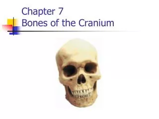
Cranium • 8 bones • Occipital • Temporal- • Sphenoid • Ethmoid • Parietal- vertex • Frontal http://www.exploringnature.org/graphics/anatomy/skulls_w_text.jpg http://www.voxel-man.de/3d-navigator/brain_and_skull/images/bs_ct-browser-englisch.jpg
Occipital Protuberance http://www.theodora.com/anatomy/images/image194.gif external internal http://img.medscape.com/pi/emed/ckb/radiology/336139-389714-1939.jpg
Temporal Bone Petrous portion • EAM
Sphenoid Bone http://img.tfd.com/MosbyMD/thumb/sphenoid_bone.jpg http://www.eurorad.org/mediafiles/eurorad/0000001070/000002_text.jpg Level of dorsum sellae= A square portion of bone on the body of the sphenoid posterior to the sella turcica or hypophysial fossa http://www.mans.eun.eg/FacMed/arabic/DAW/CourseImages/sag_mri_2za.jpg http://m.blog.hu/ke/kepalkotas/image/sella.jpg
Ethmoid Bone http://img.tfd.com/MosbyMD/thumb/ethmoid_bone.jpg Blunt Trauma- most common cause of A deviated septum Any impact can potentially detach perpendicular plate of ethmoid bone from nasal septum wall, allowing septum to deviate from one side to other.
Parietal Bone http://www.neurographics.org/3/1/2/images/Fig2.jpg http://www.cyberounds.com/assets/07/90/790/figure1.jpg
Frontal Bone http://2.bp.blogspot.com/_fBQVVpFhTQs/Sjk7DY4pljI/AAAAAAAAAvE/IYrpGnSa4rU/s320/frontal-sinus-fx-1.jpg http://www.face-and-emotion.com/dataface/anatomy/media/frontalbone.jpg The patient was admitted to the emergency room for polytrauma with severe cranioencephalic trauma with fracture and a depressed fronto-orbital fracture with external communication
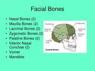
14 Facial Bones • Nasal 2x • Lacrimal 2x • Maxilla 2x • Palatine • Zygoma 2x • Inferior nasal conchae 2x • Vomer- Where is it?? • mandible http://www.asnr2.org/neurographics/8/2/60/sinusanatomy/axial/apSkull.jpg
Paranasal Sinuses • Ethmoid • Maxillary • Sphenoid • Frontal http://www.billcasselman.com/paranasal_sinuses_ethmoid_eye_sockets_sphenoid_maxillary.jpg http://scannermurcia.es/imagenes/estudios/escaner/gallery3.jpg http://www.szote.u-szeged.hu/radio/szem/fog2a1.gif
Temporomandibular Joint • Hinge of masticaction http://www.urmc.rochester.edu/smd/Rad/neuroimages/TMJ15.jpg
TMJ Open Closed http://radiopaedia.org/uploads/radio/0000/3817/TMJ_0002_gallery.jpg
TMJ-@ LPCH • CT-guided imaging at LPCH for treatment of juvenile idiopathic arthritis: The arrowshows the trajectory of the needle for placement of steroid medication into the temporomandibular joint.
Orbit http://anatomy.uams.edu/anatomyhtml/graphics/rsa2p4.gif 1. Frontal bone 2. Ethmoid bone 3. Palatine bone 4. Zygomatic bone 5. Lacrimal bone 6. Maxillary bone http://media.photobucket.com/image/anatomy%20of%20eye%20and%20muscles/trimurtulu/Eye.jpg
Orbit • What seven bones make up the orbit? • frontal, sphenoid, ethmoid (cranium) • Lacrimal, maxillary, palatine and zygomatic (facial) • The posterior obit has a what canal? • Optic- optic nerve • A blow-out fracture can cause diplopia, what does that mean? • Floor of orbit, herniation of orbital contents= doublevisiondoublevision http://e-radiography.net/ibase8/Skull/thumbs/Skull_frx_orbit_ct.jpg
Name 5 anatomic structures you can see. vertex orbit Ethmoid sinus EAM TMJ http://farm4.static.flickr.com/3122/2280819592_7a60701210.jpg
What is wrong? Rt Frontal bone- axial- orbital contents in maxillary sinus
What level is this? Petrous Ridge
What are the points at the posterior aspect? Internal/External Occipital Protuberance
What is the foreign object? What is now communicating with the external environment? Bullet Frontal Sinus open to external environment. http://www.firearmsid.com/Feature%20Articles/042002/SkullCT.jpg
What is this bone? Where is the fracture? Parietal Bone Sphenoid
Describe these two views. Pencil- Nasal cavity, ethmoid, sphenoid sinus with air/fluid (no CSF leak) http://www.powerpak.com/courses/10132/Figure3.jpg Axial- Air/ Fluid levels in the sinus http://www.ispub.com/ispub/ijorl/volume_4_number_1_37/pencil_in_the_nose/pencil-fig3.jpg
What is wrong here? Medulla and Cerebellar tonsils in foramen magnum= Chiari 1 malformation- Spina Bifida causing herniation of medulla inferior. CSF is in this space= Normal http://www.atlaschiro.com/case_studies.htm
Optic Nerve Frontal Sinus Sphenoidal Sinus Mastoid Air Cells Pons
1 • 1 Frontal Sinus • 2 Ethmoid Sinus • 3 Sphenoid Sinus • 4 Maxillary Sinus 3 2 4
2 • 1 Sphenoid Sinus • 2 Cribiform Plate 1 1 Name the structures
2 2 • 1 Maxillary sinus • 2 Frontal sinus • 3 Ethmoid sinus 3 1 1 Before Multiplanar reconstruction the 2 planes used to ACQUIRE Sinus images were AXIAL and CORONAL planes