Download
1 / 23
230 likes | 419 Views
Third Year Progress Report:. Exploiting Amyloid Fibril Lamination for Nanotube Self-Assembly. Presenter: Kun Lu Advisors: David Lynn Vince Conticello. Curr. Opinion in Struct. Biol., 2000, 10, 60-68. rod-like non-branched 8-10nm in diameter. What’s amyloid ?.

E N D
Third Year Progress Report: Exploiting Amyloid Fibril Lamination for Nanotube Self-Assembly Presenter: Kun Lu Advisors: David Lynn Vince Conticello
Curr. Opinion in Struct. Biol., 2000, 10, 60-68 rod-like non-branched 8-10nm in diameter What’s amyloid?
A(1-42),A(1-40): DAEFRHDSG10YEVHHQKLVF20FAEDVGSNKG30AIIGLMVGGV40VIA A(10-35): 10YEVHHQKLVF20FAEDVGSNKG30AIIGLM Amyloid- (A ) Protein
Solid State NMR: J. Am. Chem. Soc. 2000, 122, 7883 A(10-35) in parallel, in register orientation Small Angle Neutron Scattering (SANS) mass per unit length: 3453 340 Da/Å M. W. of A (10-35): 2855 Da distance between adjacent -strands: 5 Å one -sheet: 572 Da/Å 6 laminated -sheets
Structural Model: 10YEVHHQKLVF20FAEDVGSNKG30AIIGLM top view amplify J. Am. Soc. Chem. 2000(122):7887
only short stretches of 5-6 residues maintain H-bonding 60 ps J. Am. Chem. Soc. 2002, 124, 15150-15151 Molecular Simulation suggested fluidity of A(10-35) fibril
A(1-42) DAEFRHDSG10YEVHHQKLVF20FAEDVGSNKG30AIIGLMVGGV40VIA A(10-35) 10YEVHHQKLVF20FAEDVGSNKG30AIIGLM CH3CO-KL V F F AE-NH2 A(16-22) Designed System: E Solvent: 40% acetonitrile/Water with 0.1% TFA (pH=2.1) acidic condition: ensure amphiphilicity 40% acetonitrile: increase solubility slow down the assembly process
CD change: -sheet structure
equilibrium: at 30hr: Ribbon-like structure Uniform width: 80 5 nm Length: usually longer than 10 m Transmission Electron Microscopy (TEM)
AFM time course study: Phase image Phase image further assemble fast Large size particles ~180nm in length ~80nm in width Assembled particles Length varies no -sheet structure Round particles ~30nm by AFM no -sheet structure A (16-22) monomer Within 11hr 0-20min
Topography image Topography image Phase image Sheet twists Coil to Tubes (bent sheet) super helical ribbons -sheet structure ~90nm wide, 8nm high tubes Significant -sheet structure around 180 nm wide -sheet structure appears after 48hr Within 17hr Within 23hr
Small Angle Neutron Scattering (SANS) Small Angle X-ray Scattering (SAXS) scattering vector: Q = (4π/λ) sinθ differential neutron scattering cross-section(in a diluted system): I(q) (contrast)P(q) Contrast: difference in scattering length density between particles and solvent P(q): form factor shape, dimensions of isolated particles
hollow cylinder form factor: SAXS: SANS: outer R1=266.01 0.01 Å inner R2= 224.64 0.03 Å wall thickness= 41.4 Å outer R1=259.37 1.33 Å inner R2= 216.03 0.71 Å wall thickness= 43.3 Å
Fig. 1. Comparison of the actual fit (red curve) with the calculated scattering profile for a solid cylinder of the same outer radius (265 Å) (green curve) Fig. 2. Comparison of actual fit (red curve) with calculated scattering profiles for two hollow cylinders with the same outer radius but different wall thickness.
pitch calculation: assume they have same number of laminates Bilayer model for self-assembly of the peptide nanotubes +KL V F F A EE A F F V LK+
16-22: 10-35: N N laminates increase
absence of helical chirality AFM: right-handed: left-handed:
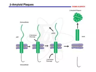
interface destabilized pH8: +KL V F F AE- - EA F F V LK+ +KL V F F AE- - EA F F V LK+ Ionized C-terminus disrupted the whole structure stable interface +KL V F F A EE A F F V LK+ pH2: +KL V F F A EEA F F V LK+
R D side chain charge Q H charge on backbone free N-E charge buried free N-Q Mutagenesis study: (no assembly) K L V F F A E free N-Q, E, G, C QK C-terminus is critical in self-assembly & N-terminus can accommodate greater diversity
Shortening A(10-35) to A(16-22) resulted in the peptide nanotube formation under designed conditions. Compared with A(10-35) fibril, the lamination order has significantly increased from 6 to 130. The resulting structures are similar to those formed by several other amphiphiles including lipids, suggesting that some intrinsic characteristic in the self-assembly process are common to various molecular frameworks. The formed nanotubes with positively charge surfaces of very different inner and outer curvature provide an easily accessible scaffold for nanotechnology. Conclusion:
Professor David G. Lynn Professor Vince P. Conticello Dr. Pappannan Thiyagarajan Dr. Jaby Jacob Dr. Robert Apkarian Argonne National Laboratory: Electron Microscopy facilities of Emory: Dr. David Morgan Dr. Ken Walsh Dr. Teresa Anne Hill Dr. Lizhi Liang Rong Gao Justin Maresh Ami S. Lakdawala Jijun Dong Peng Liu Fang fang Yan Liang Andrew G. Palmer Hsiao-Pei Liu Kaya Erbil Nora Goodman Brooke Yuri and all other conticello lab members Acknowledgement