Download
1 / 13
390 likes | 1.58k Views
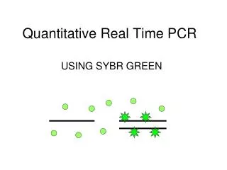
Quantitative Real-Time PCR. Adrien Six (adrien.six@pasteur.fr) Sophie Dulauroy (sophie.dulauroy@pasteur.fr) Institut Pasteur & Université Pierre et Marie Curie. Real-Time PCR: principle. Equivalent sensitivity Different Specificity.

E N D
Quantitative Real-Time PCR Adrien Six (adrien.six@pasteur.fr) Sophie Dulauroy (sophie.dulauroy@pasteur.fr) Institut Pasteur & Université Pierre et Marie Curie
Real-Time PCR: principle Equivalent sensitivity Different Specificity • The real time PCR is based on the detection and the quantification of a fluorescent transmitter during the process of amplification. • 2. The increase in the fluorescent signal is directly proportional to the quantity of amplicons produced during the reaction. • 3. Two general principles for the quantitative detection of amplicons: • - agents binding to the double-stranded-DNA (SybrGreen I) • - fluorescent probes (FAM, TAMRA, JOE, ROX,…) • For the fluorescent probes, there are 4 main technologies: • - probe hydrolysis • - hybridisation of 2 probes • - molecular beacons • - scorpion primer
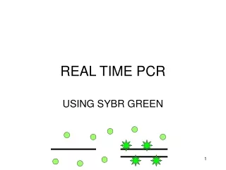
Agents binding to the double-stranded-DNA(SYBR Green I) • The free SYBR Green exhibits little fluorescence at the time of the denaturation. • With the temperature of pairing, some molecules bind to the nascent double-stranded-DNA. • During the polymerisation step, more and more of molecules bind to the nascent strand and the increase in fluorescence can be followed in real time.
Hydrolysis probes (Taqman) • During the denaturing step, the probe is free on solution. • During the annealing step, both probes hybridise to their target sequence.The proximity of the fluorochrome allows the inhibition of fluorescence. • c. The polymerase moves and hydrolyses the probe. The transmitting fluorochrome is released from the environment of the suppressor thus allowing the emission of fluorescence.
Hybridisation probes (HybProbes) • During the denaturing step, in solution the 2 probes are apart. • During the annealing step, both probes hybridise to their target sequence. The proximity of the fluorochrome allows the red emission of fluorescence. • The probes turn over free in solution.
Molecular beacons • High specificity + high precision • When T°C<Tm, hairpin structure no hybridisation and no fluorescence
Scorpion primer - Suppressor HEG prevents the replication of the molecular beacon - One of the best method
The SYBR Green: advantages and disadvantages • Advantages: • - Economic • - Easy to use • - Has more sensibility than the ethidium bromide (another intercalating agent) • - Does not inhibit the reaction of amplification • - Does not require any fluorescent probe, thus does not require any particular expertise for the design of the probes • - Is not affected by mutations in the target DNA • Disadvantages: • - Impossible to make sure of specificity of amplicons • - Bad pairing can lead to positive forgeries or an over-estimate of the quantification • - The emission of fluorescence can be skewed by the molecular mass of the DNA amplified by a longer amplicon which will fix more fluorescent molecules compared to a shorter amplicon in the same reaction • - Still unspecified mutagen capacity
Hydrolysis probes: advantages and disadvantages • Advantages: • - Increased specificity: the specificity of hybridisation between the fluorescent probe and the sequence of DNA significantly reduces the emission of non-specific fluorescence due to bad pairings or primers dimers. • - Better capacity of multiplexing: reactions multiplex can be elaborate by using distinct transmitting fluorochromes related to different probes in a PCR reaction. • Disadvantages: • - Taqman technology is less effective and less flexible device that other technologies in real time for the detection of specific mutations. • - To respect the principles of design of the probes
Hybridisation probes: advantages and disadvantages • Advantages: • High specificity • High flexibility for probe design • As the probes are not hydrolysed, they are used at each cycle • Disadvantages: • - Taqman probe design
Threshold cycle = Ct (1) • The concept of the threshold cycle is at the heart of accurate and reproducible quantification using fluorescence-based PCR. • It corresponds to the cycle from which one observes a statistically significant increase in standardized fluorescence
Threshold cycle = Ct (2) • The more template present at the beginning of the reaction, the fewer number of cycles it takes to reach a point for which the fluorescence signal is first recorded as statistically significant above background. This point is defined as the Ct. • The threshold cycle will always occur during the exponential phase of amplification.
Threshold cycle = Ct (3) • Quantification is not affected by any reaction components becoming limited in the plateau phase • The Ct value can be translated into a quantitative result by constructing a standard curve R = 2-ΔCt1/ΔCt2 ΔCt1 = ΔCt target gene = Ct target gene with treated sample – Ct same gene with sample calibrator ΔCt2 = ΔCt standardizing gene = Ct standardizing gene with treated samples – Ct same gene with sample calibrator The difference between two samples is considered as significant from one Ct.