Download
1 / 35
410 likes | 830 Views
Acute pancreatitis. The treatment of patients is dependent on the early assessment of disease severity. Potentially lethal attacks are known to occur in 2%–10% of patients with acute pancreatitis Early detect !. Classification. 1992 Atlanta, International Symposium on Acute Pancreatitis

E N D
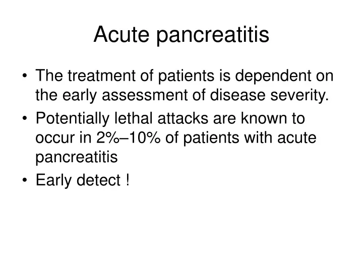
Acute pancreatitis • The treatment of patients is dependent on the early assessment of disease severity. • Potentially lethal attacks are known to occur in 2%–10% of patients with acute pancreatitis • Early detect !
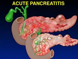
Classification • 1992 Atlanta, International Symposium on Acute Pancreatitis • Mile pancreatitis: edematous or interstitial pancreatitis, most individuals • Severe pancreatitis: necrotizing pancreatitis, occurs in about 20%–30% of all patients
Pancreatic necrosis • The importance of early detection of pancreatic necrosis in clinical practice cannot be overemphasized. • Interstitial pancreatitis: <1% mortality • Necrotizing pancreatitis: 10-23% mortality • Secondary bacterial infection: 40-70% necrotizing pancreatitis • Most patients with acute pancreatitis who develop multiorgan failure have necrotizing pancreatitis, and more than 80% of deaths occur in patients with pancreatic necrosis
How to detect? • Clinical evaluation • Laboratory investigations • Imaging studies • Ideal system • have high sensitivity and positive predictive values • be able to depict necrosis early (48 hours) • be performed rapidly (4 hours) • be available in most hospitals • be relatively inexpensive, • be objective and not observer dependent
Clinical Evaluation • Unreliable !!! • tachycardia, orthostatic hypotension, shock, respiratory distress, and signs of peritonitis: severe attack, but nonspecific, develop late • Old age, hyperlipidemia, and obesity: increased risk of death • However, these parameters are not reliable prognostic factors and cannot be used as indicators of disease severity.
Cullen's sign Turner's sign
Flank ecchymosis (Grey-Turner sign) • Periumbilical ecchymosis (Cullen sign) • More specific and have been associated with a 37% mortality rate • These signs are rarely present, however, and they often appear 48–72 hours after the onset of symptoms. • Clinical signs have only limited value for the assessment of the severity of acute pancreatitis.
Laboratory Evaluation • Amylase/Lipase: no role in the assessment of disease severity • Serum trypsinogen • Urinary trypsinogen activated peptide levels • Methemalbumin: hemorrhagic pancreatitis • Pancreatic ribonuclease: necrotic tissue • TNF • IL-6 • Phospholipase A2 • Polymophonuclear cell elastase • CRP: 2-3 days after onset, low specific
Multiple prognostic indexes • Reflect systemic abnormalities • Not correlate well with severity and extent of local disease • Ranson scores: corresponding increase in the morbidity and mortality rates. • <3 : no mortality • >6 : mortality rate >50%, usually necrotizing pancreatitis • a total of 48 hours of observation for proper evaluation.
Multiple prognostic indexes • Acute Physiology and Chronic Health Evaluation (APACHE II) • Corresponding increase in morbidity and mortality • >8: severe pancreatitis • Advantage: used throughout the patient’s hospital course in monitoring the patient’s response to therapy
Imaging study • Conventional abdominal radiographs and barium studies: no role in the early evaluation of disease severity. • CXR ±renal function tests: useful for the prediction of severity • pulmonary findings (infiltrates, effusions) in acute pancreatitis is 15%–55%, seen mainly in patients with severe disease. • The predictive value is increased with left-sided or bilateral pleural effusions.
Ultrasonographic Evaluation • Indicated early in an acute episode of pancreatitis • help evaluate gallbladder and/or common duct stones. • Interfered with bowel gas • Intraparenchymal and retroperitoneal fluid collections correlates poorly with pancreatic necrosis. • A diffusely enlarged and hypoechoic gland is consistent with interstitial edema, while extrapancreatic fluid collections (lesser sac, anterior pararenal space) are detected in patients with severe disease
CT Evaluation • Contrast material–enhanced CT • quantify pancreatic parenchymal injury • mild pancreatitis have an intact capillary network
Areas of diminished or no enhancement indicate decreased blood flow and relate to pancreatic zones of ischemia or necrosis • CT has shown an overall accuracy of 87%with a sensitivity of 100% for the detection of extended pancreatic necrosis and a sensitivity of 50% if only minor necrotic areas were present at surgery.
Without necrosis: no mortality and a complication rate of only 6% • CT evidence of necrosis: mortality 23%, morbidity 82%.
<30% necrosis: no mortality and a 48% morbidity rate, • >30%: mortality rate of 11–25%, morbidity 75%–100% • It should be emphasized, however, that systemic and local complications may occur during an episode of acute pancreatitis in patients without pancreatic necrosis.
CT Enhancement Values and Pitfalls • Fatty infiltration of the pancreas • Interstitial pancreatitis, due to parenchymal edema. • Small intrapancreatic fluid collections • The distinction is difficult and sometimes impossible to make unless previous or follow-up CT images are available for review.
Pancreatic necrosis develops early, within the first 24–48 hours after the onset • zones of tissue liquefaction become better defined and more easily recognized 2–3 days after the initial onset • Thus CT scans obtained 3 days after clinical onset yield higher accuracy in the depiction of necrotizing pancreatitis
Fat necrosis: occurs in patients with or without parenchymal necrosis • Favor secondary bacterial contamination. • 22% incidence of local complications in patients without pancreatic necrosis but with peripancreatic fluid collections • CT cannot be used to help reliably diagnose, nor can it help accurately quantify, retroperitoneal fat necrosis. • It has been suggested, therefore, that in clinical practice all heterogeneous peripancreatic collections should be considered areas of fat necrosis until proven otherwise
CT Correlation with NumericSystems • There is a wide variation in the relationship of prognostic signs (Ranson, APACHE II) and early CT findings in acute pancreatitis. • CT evaluation results were found to be better prognostic indicators, owing to greater sensitivity and specificity.
Magnetic Resonance Imaging • an excellent alternative noninvasive modality to help evaluate patients and stage acute pancreatitis • particularly useful in patients who cannot receive iodinated contrast material due to allergic reactions or renal insufficiency. • Gadolinium-enhanced T1-weighted: pancreatic necrosis as areas of nonenhanced parenchyma • Fat-suppression images: defining subtle, diffuse, or focal parenchymal abnormalities. • T2-weighted images: Fluid collections, pseudocysts, and areas of hemorrhage.
CONCLUSIONS • Numeric systems (APACHE II, Ranson) are commonly used today to help detect organ failure, and the acquired data are used as indirect evidence of disease severity, with a sensitivity of about 70%. • Contrast-enhanced CT is the imaging modality of choice to help stage the severity of inflammatory processes, detect pancreatic necrosis, and depict local complications.