Download
1 / 60
680 likes | 998 Views
Acute Pancreatitis. Steven B. Goldin, MD University of South Florida. J.H. JH is a 64-yr-old male admitted to an outside hospital with a 4 day history of progressively worsening epigastric pain without radiation. History. What other points of the history do you want to know?.

E N D
Acute Pancreatitis Steven B. Goldin, MD University of South Florida
J.H. JH is a 64-yr-old male admitted to an outside hospital with a 4 day history of progressively worsening epigastric pain without radiation.
History What other points of the history do you want to know?
History, J.H. Consider the Following • Characterization of symptoms • Temporal sequence • Alleviating / Exacerbating factors: • Pertinent PMH, ROS, MEDS. • Relevant family hx. • Associated signs and symptoms
HPI J.H. Pain is constant and unremitting, going through to his back Pain started after beer and pizza 4 days prior, progressively worsening since Nausea and vomiting x 3 Some indigestion history, never like this No relief with OTC Pepcid, Mylanta or Advil
History, J.H. No significant past medical or surgical history No medications No allergies to medications Smokes 1ppd x 40 yrs, and drinks ethanol heavily. He denies drug use. Family history was noncontributory.
Differential DiagnosisBased on History and Presentation Cholecystitis Choledocholithiasis PUD Gastritis Pancreatitis Bowel obstruction Mesenteric ischemia Gastroenteritis Appendicitis Hepatitis Diabetes Rectus hematoma Pneumonia Pyelonephritis Trauma w/ duodenal hematoma.
Physical Examination What would you look for on physical examination?
Physical Examination, J.H. Vital Signs: T 38.5 BP 120/70 P 100 R18 Appearance: lying still in moderate distress. Not jaundiced and sclera were anicteric. His mucous membranes were dry. Resp: His lungs were clear to auscultation. CV: heart was regular and without murmurs, rubs, or gallops.
Physical Examination, J.H. Abdomen: soft, moderately distended, tender in the mid-epigastric region and right upper quadrant. No palpable masses. Bowel sounds were positive. Extremities: without cyanosis, clubbing, or edema. Rectal exam: no masses, guaiac neg.
Revised Differential Cholecystitis PUD Pancreatitis Bowel obstruction Mesenteric ischemia Gastroenteritis Hepatitis Rectus hematoma Pneumonia
Laboratory What would you obtain?
LabsConsider the following CBC, Electrolytes, LFT’s, CMP, LDH, Amylase, Lipase, PT, PTT, Urinalysis, ABG,
Lab Results, Discussion This patient has a hypokalemic hypochloremic metabolic alkalosis from vomiting. He has an elevated amylase and lipase consistent with pancreatitis. On admission he has 4 out of 5 of Ranson’s criteria and can be expected to become very sick. There are 6 more of Ranson’s criteria that should be tracked over the next 48 hours. Can you list Ranson’s criteria?
Ranson’s Early Objective Prognostic Signs that Correlate with the Risk of Major Complications or Death On Admission Non-biliary Biliary Age >55 >70 WBC >16 >18 Glucose >200 >220 LDH >350 >400 SGOT >250 >250
Ranson’s Early Objective Prognostic Signs that Correlate with the Risk of Major Complications or Death During the Initial 48 Hours Non-biliary Biliary Hematocrit decrease >10% >10% BUN increase >5 mg/dL >2 mg/dL Calcium <8 mg/dL <8 mg/dL Arterial Po2 <60 ... Base deficit >4 mEq/L >5 mEq/L Fluid sequestration >6 L >4 L
Ranson’s Prognostic Signs that Correlate with the Risk of Major Complications or Death
Ranson’s Early Objective Prognostic Signs that Correlate with the Risk of Major Complications or Death Note 1. The amylase and lipase levels are not prognostic signs and do not relate to the severity of the attack or prognosis. 2. LDH must usually be specifically ordered. It is not included with most comprehensive metabolic panels or with most liver function tests.
Interventions at this point IVF – LR Bolus 1-2 liters then LR at 150cc/hr – titrate to urine output/volume status NPO Foley catheter NG Tube Admission to ICU
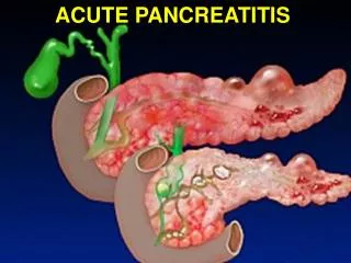
Pancreatitis Alcohol * Gallstones* Hyperlipidemia Trauma Tumor Ischemia Medications Infection Post-op/Post-procedure Other Idiopathic
Studies What would you order ?
Discussion of Studies Ultrasound of right upper quadrant is indicated to evaluate gallbladder and bile duct for stones. CT scan should be done after initial stabilization. IV contrast is useful to assess pancreatic viability. Use of IV contrast on presentation is debated.
Discussion of imaging study This is a CT scan of the abdomen done with both oral and IV contrast. It demonstrates edema surrounding the pancreas and is consistent with the laboratory results suggesting pancreatitis. No significant pancreatic necrosis is noted. My preference is to not use IV contrast on admission if pancreatitis is suspected due to the toxic nature of the dye and the rarity of finding infection on presentation. I do use IV contrast later on in the hospitalization to better discern the amount of necrosis that has resulted as long as the patients renal function is acceptable.
Revised Differential Diagnosis Acute Pancreatitis Choledocholithiasis Cholecystitis Perforated ulcer
Supportive measures Nothing by mouth early oral feedings may increase the severity of pancreatic inflammation. Oral feedings should be withheld until resolution of abdominal pain, fever, and leukocytosis Fluid and electrolyte repletion and resuscitation Respiratory support Nutritional support Proton pump inhibitors DVT prophylaxis Antibiotics (debated) Analgesics
Timing of cholecystectomy Gallstones are present in 60% of non-alcoholic patients with pancreatitis and if allowed to persist, 36 - 63% will develop recurrent bouts of pancreatitis. Cholecystectomy reduces this risk to 2 - 8%.
Timing of cholecystectomy 75% of patients with acute abdominal pain, gallstones, and elevated amylase have no gross evidence of significant pancreatitis. Cholecystectomy is safe in this group. In patients with gross evidence of pancreatitis, 80% have mild disease and cholecystectomy is safe but does not alter the course of the pancreatitis
Intra-Operative Cholagiogram (IOC) during Laparoscopic Cholecystectomy
The timing of cholecystectomy In patients with severe pancreatitis there is an 82.6% morbidity and 47.8% mortality from cholecystectomy if performed within the initial 48 hours. If deferred until the signs of pancreatitis have subsided, morbidity and mortality fall to 17.8% and 11.8% respectively.
Timing of cholecystectomy In patients with severe pancreatitis and an obstructed biliary tree secondary to choledocholithiasis, ERCP and sphincterotomy significantly reduce morbidity related to biliary complications but do not alter the course of the pancreatic inflammation.
Hospital Course This patient deteriorates with non-operative treatment. He develops high fevers and hypotension. What could be happening? What would you do next?
Repeat CT Scan is shown below What do you see?
CT Findings CT scan now shows air in the lesser sac. This is diagnostic of infected pancreatic necrosis. What next?
What next? Supportive Treatment Elective Cholecystectomy if caused by gallstones. Endoscopy with ERCP if obstructing stone is identified in the common bile duct. OR if infected
Management The patient was started on broad spectrum antibiotics and taken to the operating room for pancreatic debridement, cholecystectomy, and placement of large axiom sump drains. A jejunal feeding tube was also placed at this time.
Temporary Abdominal Closure Sump Drain
Management This patient was slowly weaned from his vasopressor agents and ventilator. Tube feedings were started two days after his debridement. The patient eventually made a full recovery and was discharged from the hospital approximately 4 months after presentation.
Management Operative Options include Debridement and drainage-Mortality 13.9%. 58.3% of patients can be treated with one surgical procedure. Debridement & packing, and dressing changes every 2 - 3 days. Mortality 10.7%. My preference is to debride and drain if all necrotic debris can be easily removed. Otherwise I pack and return to the operating room every 48 hours until the necrotic tissue is fully debrided. At that time, I place drains and close the patient.
Management Patients who are not infected should not be operated on. Bradley - Neither the existence nor the extent of necrosis can be used as an indication for surgery. (90.4% survival in patients treated conservatively with over 50% necrosis of the gland and no infection).