Download
1 / 28
280 likes | 389 Views
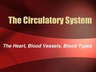
UNIT 2 THE CIRCULATORY SYSTEM CHAPTER 12 WELCOME! JULIE J. MCDONALD, BS, MPT. TODAY’S AGENDA. Welcome Course Questions, Problems & Issues Seminar discussion. Course Questions, Problems & Issues. Unit 1 Issues to Review. Universal Donor: Type O—no A or B antigens present on cells

E N D
UNIT 2THE CIRCULATORY SYSTEMCHAPTER 12WELCOME!JULIE J. MCDONALD, BS, MPT
TODAY’S AGENDA • Welcome • Course Questions, Problems & Issues • Seminar discussion
Unit 1 Issues to Review • Universal Donor: Type O—no A or B antigens present on cells • Universal Recipient: Type AB—no A, B or O antibodies present in plasma • Erythroblastosis fetalis: Rh- mom has second Rh+ baby; mom’s Rh antibodies attack baby’s Rh+ cells; prevented by giving Mom RhoGam after first delivery
Unit 1 Issues to Review • Hemoglobin: A chemical pigment in the RBCs that traps O2; made up of protein chains and Fe+ atoms • WBCs: Differential count measures proportions of each type of WBC in blood sample • Phagocytes: Neutrophils most numerous; Eosinophils weak; Monocytes most agressive
Unit 1 Issues to Review • Anticoagulants: Coumadin, Heparin (NOT Vitamin K) • Embolus: Circulating blood clot (a thrombus that has broken free)
CHAPTER 12 THE CIRCULATORY SYSTEM • Heart • Blood Vessels • Circulation • Blood Pressure
Location, Size, and Position of the Heart • In mediastinum • 2/3 to the left of the body midline • Apex = point • Most inferior portion • Shape and size of a closed fist • Septum divides right and left sides (internally)
Anatomy of the Heart • Heart chambers • Upper • Right and left atria (atrium) • Small chambers • Receiving Chambers • Lower • Right and left ventricles • Larger chambers • Discharging Chambers
SUMMARY OF LAYERS OF THE HEART • Outside (external) to Inside (internal) • Parietal Pericardium • Pericardial cavity (filled with fluid) • Visceral Pericardium/Epicardium • Myocardium • Endocardium
Three layers of the Heart Wall • Epicardium • Outer layer • Connective tissue • Myocardium • Middle layer • Thick • Muscle (My/o) • Endocardium • Inner layer (lining) • Very thin, smooth
Coronary Circulation • Blood for the myocardium of the heart, flows through the right and left coronary arteries • Blockage of blood flow through the coronary arteries can cause myocardial infarction (heart attack)
Vessels • Pulmonary Arteries • Carry de-oxygenated blood from R ventricle to lungs • R pulmonary artery to R lung • L pulmonary artery to L lung • Pulmonary Veins • Carry oxygenated blood from lungs to L atria • R pulmonary veins from R lung • L pulmonary veins from L lung
VESSELS (CONT.) • Vena Cava • Largest Veins • Inferior (IVC) and Superior (SVC) • Empties blood into R atrium from systemic circulation
Valves • Cuspid valves • Tricuspid: between right atrium and ventricle • Bicuspid (mitral): between left atrium and ventricle • Open and close from chordae tendineae • Semilunar valves • Pulmonary Semilunar: base of pulmonary arteries • Aortic Semilunar: base of aorta • Open and close from pressure within heart
The heart acts as two pumps Right atrium and ventricle • pump deoxygenated blood to the lungs • “Pulmonary Circulation” Left atrium and ventricle • pump oxygenated blood to the body • “Systemic Circulation
Blood Flow Pathway ➥Right atrium ➥Tricuspid valve ➥Right ventricle ➥Pulmonary Semilunar Valve ➥Pulmonary Arteries ➥Lungs ➡ ➥Pulmonary Veins ➥Left atrium ➥Bicuspid valve ➥Left Ventricle ➥Aortic Semilunar Valve ➥Aorta ➥Arterioles ➥Capillaries➡O₂/CO₂ exchange
The Heart—Actions • Relaxation: Diastole • Contraction: Systole
Conduction System of the Heart • SA (sinoatrial) node • The pacemaker • In wall of right atrium near superior vena cava • AV (atrioventricular) node • In the floor of right atrium near septum • AV bundle (bundle of His) • Located in the septum of the ventricle • Purkinje fibers— • Located in the walls of the ventricles • Cause contraction of myocardium • Coordination of impulses cause atrial contraction f/b ventricular contraction…
Conduction System of the Heart • The normal ECG has three deflections or waves called the P wave, the QRS complex, and the T wave • P wave—associated with depolarization of the atria • QRS complex—associated with depolarization of the ventricles • T wave—associated with repolarization of the ventricles
Heart Sounds • Two distinct heart sounds in every heartbeat or cycle—“lubb-dupp” • First (lubb) sound is caused by the vibration and closure of AV valves during contraction of the ventricles • Second (dupp) sound is caused by the closure of the semilunar valves during relaxation of the ventricles
Cardiac Cycle • Heart beat is regular and rhythmic—each complete beat called a cardiac cycle—average is about 72 beats per minute • Each cycle, about 0.8 seconds long, subdivided into systole (contraction phase) and diastole (relaxation phase)
Cardiac Cycle • Stroke volume is the volume of blood ejected from one ventricle with each beat • Cardiac output is amount of blood that one ventricle can pump each minute—average is about 5 L per minute at rest • Blood Pressure= measures the gradient between pressure at the aorta and vena cava • MABP= CO x SV
SOME OTHER ODDS AND ENDS… Assignment– Unit 4 Unit 3- Lymphatic System and Immunity