Download
1 / 34
350 likes | 559 Views
Proteins Function and Structure. Proteins. more than 50% of dry mass of most cells functions include structural support storage, transport cellular communication movement defense against foreign substances (immunity) - enzymatic reactions. Cell communication.

E N D
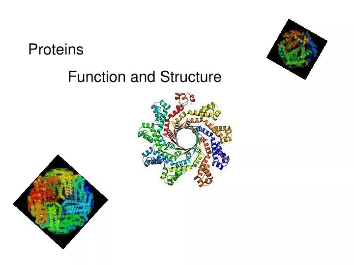
Proteins Function and Structure
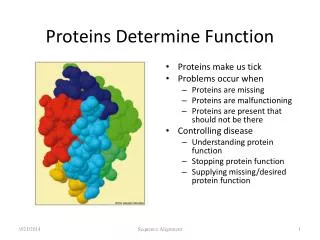
Proteins • more than 50% of dry mass of most cells • functions include • structural support • storage, transport • cellular communication • movement • defense against foreign substances (immunity) - enzymatic reactions
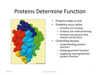
Think of conditions or diseases where malfunctioning proteins are responsible?
Structure of Proteins • Monomer: amino acid • 20 different a.a. used in cells • Polymer of amino acids-->polypeptide Complex of >1 polypeptides-->protein (a protein often refers to the functional entity)
Amino Acid Structure • Organic molecules with • Amino end ? • Carboxyl end ? • Central -carbon • Distinct side chain (or R group) bonded to -carbon
a carbon LE 5-UN78 H+ What happens to the end groups in a cellular environment? Carboxyl group Amino group
How are 20 amino acids be different from each other? R groups are unique Note: R groups- aka side chains
Glycine (Gly) Amino acids Memorize structure LE 5-17a Alanine (Ala) Valine (Val) Isoleucine (Ile) Leucine (Leu) Nonpolar Methionine (Met) Phenylalanine (Phe) Proline (Pro) Tryptophan (Trp)
LE 5-17b Polar Tyrosine (Tyr) Serine (Ser) Asparagine (Asn) Threonine (Thr) Cysteine (Cys) Glutamine (Gln)
LE 5-17c Acidic Basic Electrically charged Aspartic acid (Asp) Lysine (Lys) Arginine (Arg) Glutamic acid (Glu) Histidine (His)
Amino acids • linked together through peptide bonds • Draw dipeptide bond showing bond • Polypeptides range in length • a few a.a. to > thousand • Each polypeptide has unique linear sequence of amino acids
Protein Conformation • Helices, coils, pleats • Sequence of amino acids determines 3-D conformation--> function • Depicted in ribbon and space-filling models
LE 5-19 Groove A ribbon model Groove A space-filling model
Four Levels of Protein Structure • Primary structure (1o) • unique sequence of amino acids, like letters in a word • Secondary structure (2o) • -helices and -pleated sheets • Stabilized by H-bonds • Tertiary structure (3o) • determined by interactions among various side chains (R groups) • Quaternary structure (4o) • Multiple polypeptide chains forming a functional protein
Amino end Amino acid subunits 1o structure LE 5-20a Gly Ser Tyr Phe…. Carboxyl end
Four Levels of Protein Structure • Primary structure (1o) • unique sequence of amino acids, like letters in a word • Secondary structure (2o) • -helices and -pleated sheets • Stabilized by H-bonds between amino and carbonyl groups - Creates 3-D conformation • Tertiary structure (3o) • determined by interactions among various side chains (R groups) • Quaternary structure (4o) • Multiple polypeptide chains forming a functional protein
2o structure LE 5-20b b pleated sheet Amino acid subunits helix
Four Levels of Protein Structure • Primary structure (1o) • unique sequence of amino acids, like letters in a word • Secondary structure (2o) • -helices and -pleated sheets • Stabilized by H-bonds • Tertiary structure (3o) - determined by bonds between side chains (R groups) often between linearly distant amino acids -ionic bonds, disulfide bonds, van der Waals forces, H-bonds - creates to 3-D conformation • Quaternary structure (4o) • Multiple polypeptide chains forming a functional protein
LE 5-20d Hydrophobic interactions and van der Waals interactions Polypeptide backbone Hydrogen bond Cysteines form disulfide bonds. Look at R group of cysteine to see why. Disulfide bridge Ionic bond
Four Levels of Protein Structure • Primary structure (1o) • unique sequence of amino acids, like letters in a word • Secondary structure (2o) • -helices and -pleated sheets • Stabilized by H-bonds • Tertiary structure (3o) - determined by bonds between side chains (R groups) often between linearly distant amino acids -ionic bonds, disulfide bonds, van der Waals forces, H-bonds - contributes to 3-D conformation • Quaternary structure (4o) • Multiple polypeptide chains forming a functional protein
How many of you have or are singing in a choir? How many in the group? Play on a team? How many on the team? Worked in theater? With how many others? Could you have accomplished the group’s goal alone?
Polypeptide chain LE 5-20e b Chains Iron Heme Complex of polypeptide subunits a Chains Hemoglobin Collagen
LE 5-20 b pleated sheet +H3N Amino end Amino acid subunits helix
Significance of Protein Conformation • Small change in 1o structure • can change protein’s conformation and function • Example • Sickle-cell disease • an inherited blood disorder-->anemia Caused by single amino acid substitution in hemoglobin
Sickled RBC Normal RBC 10 µm 10 µm Fibers of abnormal hemoglobin deform cell into sickle shape. Normal cells are full of individual hemoglobin molecules, each carrying oxygen. LE 5-21a
One Amino Acid Substitution: Huge Effect! Sickle-cell hemoglobin Normal hemoglobin Primary structure Primary structure Val Val His His Thr Pro Glu Glu Thr Pro Val Glu Leu Leu LE 5-21b 1 1 2 4 6 2 4 6 7 7 3 5 3 5 Exposed hydrophobic region Secondary and tertiary structures Secondary and tertiary structures b subunit b subunit a a Quaternary structure Sickle-cell hemoglobin Normal hemoglobin (top view) Quaternary structure a a Function Molecules do not associate with one another; each carries oxygen. Molecules interact with one another to crystallize into a fiber; capacity to carry oxygen is greatly reduced. Function
Environment Affects Protein Structure & Function ? pH salt concentration temperature other environmental factors Extreme conditions cause unraveling of protein structure:denaturation
Caused by, for example, high temperature (100oC) Denaturation LE 5-22 Normal protein Denatured protein Renaturation Lowered Temp (37oC)
Proper Protein-Folding • Chaperonins • protein complexes that assist in the proper folding of other proteins
Cap LE 5-23a Hollow cylinder Chaperonin (fully assembled)
Model Correctly folded protein LE 5-23b Polypeptide Steps of Chaperonin Action: The cap attaches, causing the cylinder to change shape in such a way that it creates a hydrophilic environment for the folding of the polypeptide. The cap comes off, and the properly folded protein is released. An unfolded poly- peptide enters the cylinder from one end.
Techniques to Determine Protein Structure • X-ray crystallography (need to make protein crystals) • Nuclear magnetic resonance (NMR) spectroscopy (not dependent on making protein crystals)
How is the sequence of proteins determined? -encoded in DNA - two step process to decode DNA is transcribed into mRNA 2. mRNA is translated into polypetide More later