Download
1 / 53
570 likes | 960 Views
The Structure and Function of Nucleic Acids and Proteins Macromolecules. Macromolecules Are large molecules composed of smaller molecules Are complex in their structures. Concept 1: Most macromolecules are polymers, built from monomers.

E N D
The Structure and Function of Nucleic Acids and Proteins Macromolecules
Macromolecules • Are large molecules composed of smaller molecules • Are complex in their structures
Concept 1: Most macromolecules are polymers, built from monomers Three of the classes of life’s organic molecules are polymers • Carbohydrates • Proteins • Nucleic acids
1 HO H 3 2 H HO Unlinked monomer Short polymer Dehydration removes a watermolecule, forming a new bond H2O 1 2 3 4 HO H Longer polymer (a) Dehydration reaction in the synthesis of a polymer The Synthesis and Breakdown of Polymers • A polymer is a long molecule consisting of many similar building blocks called monomers • Monomers form larger molecules by condensation reactions called dehydration reactions
1 3 HO 4 2 H Hydrolysis adds a watermolecule, breaking a bond H2O 1 2 H HO 3 H HO (b) Hydrolysis of a polymer • Polymers can disassemble by • Hydrolysis
The Diversity of Polymers • Each class of polymer • Is formed from a specific set of monomers • Although organisms share the same limited number of monomer types, each organism is unique based on the arrangement of monomers into polymers • An immense variety of polymers can be built from a small set of monomers 1 3 2 H HO
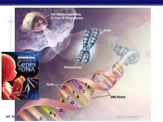
Concept 2: Nucleic acids store and transmit hereditary information • Genes • Are the units of inheritance • Program the amino acid sequence of polypeptides • Are made of nucleic acids
The Roles of Nucleic Acids • There are two types of nucleic acids • Deoxyribonucleic acid (DNA) • Ribonucleic acid (RNA) • DNA • Stores information for the synthesis of specific proteins
DNA 1 Synthesis of mRNA in the nucleus mRNA NUCLEUS CYTOPLASM mRNA 2 Movement of mRNA into cytoplasm via nuclear pore Ribosome 3 Synthesis of protein Aminoacids Polypeptide • Directs RNA synthesis • Directs protein synthesis through RNA
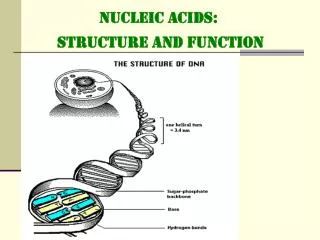
5’ end 5’C O 3’C O O 5’C O 3’C 3’ end OH The Structure of Nucleic Acids • Nucleic acids • Exist as polymers called polynucleotides (a) Polynucleotide, or nucleic acid
Nucleoside Nitrogenous base O 5’C O O CH2 P O O Phosphate group 3’C Pentose sugar (b) Nucleotide • Each polynucleotide • Consists of monomers called nucleotides
Nitrogenous bases Pyrimidines NH2 O O C C CH3 C N CH HN C CH HN CH CH C CH C C CH CH N N O N O O H H H Cytosine C Uracil (in RNA) U Thymine (in DNA) T Uracil (in RNA) U Purines O NH2 C C N N C C NH N HC HC C CH C N N NH2 N N H H Adenine A Guanine G Pentose sugars 5” 5” OH OH HOCH2 HOCH2 O O H H H H 1’ 1’ 4’ 4’ H H H H 3’ 2’ 3’ 2’ H OH OH OH Deoxyribose (in DNA) Ribose (in RNA) Ribose (in RNA) Nucleotide Monomers • Nucleotide monomers • Are made up of nucleosides and phosphate groups Pyrimidines Purines (c) Nucleoside components
Nucleotide Polymers • Nucleotide polymers • Are made up of nucleotides linked by the–OH group on the 3´ carbon of one nucleotide and the phosphate on the 5´ carbon on the next • The sequence of bases along a nucleotide polymer • Is unique for each gene
The DNA Double Helix • Cellular DNA molecules • Have two polynucleotides that spiral around an imaginary axis • Form a double helix
3’ end 5’ end Sugar-phosphatebackbone Base pair (joined byhydrogen bonding) Old strands Nucleotideabout to be added to a new strand 3’ end A 5’ end Newstrands 3’ end 3’ end 5’ end • The DNA double helix • Consists of two antiparallel nucleotide strands
Nucleoside Nitrogenous base O 5’C O O CH2 P O O Phosphate group 3’C Pentose sugar (b) Nucleotide • The nitrogenous bases in DNA • Form hydrogen bonds in a complementary fashion (A with T only, and C with G only)
James Watson and Francis Crick with a model of the DNA molecule, the double helix.
X-Ray Diffraction Photograph of a Hydrated DNA Fiber Rosalind Franklin: The Dark Lady of DNA (Brenda Maddox, 2002) Photograph 51 Dr. Rosalind Franklin
討論: • DNA有何特徵? • 1 • 2 • 3
討論: • RNA有何特徵? • 1 • 2 • 3
Concept 3: Proteins have many structures, resulting in a wide range of functions • Proteins • Have many roles inside the cell
Substrate binds to enzyme. 1 Active site is available for a molecule of substrate, the reactant on which the enzyme acts. 2 2 Substrate (sucrose) Glucose Enzyme (sucrase) OH H2O Fructose H O 4 Products are released. 3 Substrate is converted to products. • Enzymes • Are a type of protein that acts as a catalyst, speeding up chemical reactions
Polypeptides • Polypeptides • Are polymers of amino acids • A protein • Consists of one or more polypeptides
Amino Acid Monomers • Amino acids • Are organic molecules possessing both carboxyl and amino groups • Differ in their properties due to differing side chains, called R groups
CH3 CH3 CH3 CH CH2 CH3 CH3 H CH3 H3C CH3 CH2 CH O O O O O H3N+ C H3N+ C H3N+ H3N+ C C C C C C H3N+ C C O– O– O– O– O– H H H H H Valine (Val) Leucine (Leu) Isoleucine (Ile) Glycine (Gly) Alanine (Ala) Nonpolar CH3 CH2 S H2C CH2 O NH CH2 C C H2N CH2 CH2 O– CH2 O O O H H3N+ H3N+ C C C C H3N+ C C O– O– O– H H H Phenylalanine (Phe) Proline (Pro) Methionine (Met) Tryptophan (Trp) • 20 different amino acids make up proteins
OH NH2 O C NH2 O C OH SH CH2 CH3 OH Polar CH2 CH CH2 CH2 CH2 CH2 O O O O O O H3N+ C H3N+ C H3N+ C C H3N+ C C H3N+ C C C C C H3N+ C O– O– O– O– O– O– H H H H H H Glutamine (Gln) Tyrosine (Tyr) Asparagine (Asn) Cysteine (Cys) Serine (Ser) Threonine (Thr) Basic Acidic NH3+ NH2 NH+ O– O –O O CH2 C NH2+ C C NH Electrically charged CH2 CH2 CH2 CH2 CH2 O O CH2 CH2 C CH2 C H3N+ C H3N+ C O O– O– CH2 C H3N+ CH2 C H O H O– C C H3N+ CH2 H O O– C C H3N+ H O– H Lysine (Lys) Histidine (His) Arginine (Arg) Glutamic acid (Glu) Aspartic acid (Asp)
Peptidebond OH SH CH2 CH2 CH2 H H H C C H C C N C OH H C OH N N DESMOSOMES H O H O H O (a) H2O OH DESMOSOMES DESMOSOMES Side chains SH OH Peptidebond CH2 CH2 CH2 H H H N OH C C C C C H C N N Backbone H H O O H O Amino end(N-terminus) Carboxyl end(C-terminus) (b) Amino Acid Polymers • Amino acids • Are linked by peptide bonds OH
Determining the Amino Acid Sequence of a Polypeptide • The amino acid sequences of polypeptides • Were first determined using chemical means • Can now be determined by automated machines
Groove (a) A ribbon model Groove (b) A space-filling model Protein Conformation and Function • A protein’s specific conformation • Determines how it functions • Two models of protein • conformation:
+H3NAmino end Pro Thr Gly Gly Amino acid subunits Thr Gly Glu Seu Lys Cys Pro Leu Met Val Lys Val Leu Asp Ala Arg Val Gly Ser Pro Ala Glu Lle Asp Thr Lys Ser Tyr Trp Lys Ala Leu Gly lle Ser Pro Phe His Glu His Ala Glu Val Thr Phe Val Ala Asn lle Thr Asp Ala Tyr Arg Ser Ala Arg Pro Gly Leu Leu Ser Pro Tyr Ser Tyr Ser Thr Thr Ala o Val c Val Glu Lys o Thr – Pro Asn Carboxyl end Four Levels of Protein Structure • Primary structure • Is the unique sequence of amino acids in a polypeptide
H H H H H H O O O O O O O H H H H H H R R R R R R R C C C C C C C C C C C C C N N N N N N N N N N N N N C C C C C C C C C C C C C C R R R R R R H H H H H H H O O O O O O O H H H H H H H pleated sheet H O H H Amino acidsubunits C C N N N C C C R H O H H H H H H N N N N N N helix C C O C H H H C C C R R R R R H H C C C C C C O O O O H C R O C C O H C O N N H C C H R H R • Secondary structure • Is the folding or coiling of the polypeptide into a repeating configuration • Includes the helix and the pleated sheet
Hydrophobic interactions and van der Waalsinteractions CH CH2 CH2 H3C CH3 OH Polypeptidebackbone H3C CH3 Hyrdogenbond CH O HO C CH2 CH2 S S CH2 Disulfide bridge O -O C CH2 CH2 NH3+ Ionic bond • Tertiary structure • Is the overall three-dimensional shape of a polypeptide • Results from interactions between amino acids and R groups
Polypeptidechain Collagen Chains Iron Heme Chains Hemoglobin • Quaternary structure • Is the overall protein structure that results from the aggregation of two or more polypeptide subunits
+H3N Amino end Amino acid subunits helix • The four levels of protein structure
Sickle-Cell Disease: A Simple Change in Primary Structure • Sickle-cell disease • Results from a single amino acid substitution in the protein hemoglobin
Normal hemoglobin Sickle-cell hemoglobin Primary structure Primary structure . . . . . . Exposed hydrophobic region Val His Leu Thr Pro Glul Glu Val His Leu Pro Glu Thr Val 5 6 7 3 4 5 6 7 1 2 1 2 3 4 Secondaryand tertiarystructures Secondaryand tertiarystructures subunit subunit Quaternary structure Hemoglobin A Quaternary structure Hemoglobin S Molecules interact with one another tocrystallize into a fiber, capacity to carry oxygen is greatly reduced. Function Molecules donot associatewith oneanother, eachcarries oxygen. Function 10 m 10 m Normal cells arefull of individualhemoglobinmolecules, eachcarrying oxygen Red bloodcell shape Red bloodcell shape • Hemoglobin structure and sickle-cell disease Fibers of abnormalhemoglobin deform cell into sickle shape.
What Determines Protein Conformation? • Protein conformation • Depends on the physical and chemical conditions of the protein’s environment
Denaturation Normal protein Denatured protein Renaturation Denaturation • Is when a protein unravels and loses its native conformation
The Protein-Folding Problem • Most proteins • Probably go through several intermediate states on their way to a stable conformation
Correctlyfoldedprotein Polypeptide Cap Hollowcylinder The cap attaches, causing the cylinder to change shape insuch a way that it creates a hydrophilic environment for the folding of the polypeptide. The cap comesoff, and the properlyfolded protein is released. Chaperonin(fully assembled) Steps of ChaperoninAction: An unfolded poly- peptide enters the cylinder from one end. 2 1 3 Chaperonins • Are protein molecules that assist in the proper folding of other proteins