Download
1 / 17
170 likes | 334 Views
Semi-automated Archiving of Scanned Requisition Documents in Anatomic Pathology. Document Imaging. Electronically imaging documents yields many of the same advantages as “going digital” does for images Accessibility Availability Storage. Requisition Handling Issues.

E N D
Semi-automated Archiving of Scanned Requisition Documents in Anatomic Pathology
Document Imaging • Electronically imaging documents yields many of the same advantages as “going digital” does for images • Accessibility • Availability • Storage John H. Sinard, MD, PhD
Requisition Handling Issues • Sometimes contain useful clinical information • Would never “embed” in a report, so no “need” to import into LIS • Desirable to have available at time of signout • Frequent sorting • Primary copy sorted to allow collating with working drafts • Gross room copy sorted for filing • Billing copy transferred to Accounting service for Registration • Sorted after signout for filing • Removed from working drafts and sent for microfilming • Need to be retained for minimum of 2 years John H. Sinard, MD, PhD
Requisition Costs • With >90,000 cytology specimens and >45,000 surgical specimens per year, represents about 150,000 unique documents, most of which are copied at least once to support our workflow • Department was spending $15-25K per year for microfilming or contracted digital archiving services • The major cost is man-hours spent sorting, copying, collating with working drafts, distributing, resorting, removing from working drafts, removing staples, resorting, sending for microfilming • Minimum of 5-10 hrs / day department-wide, or approximately 1 FTE • Does NOT include time spent looking for/retrieving documents John H. Sinard, MD, PhD
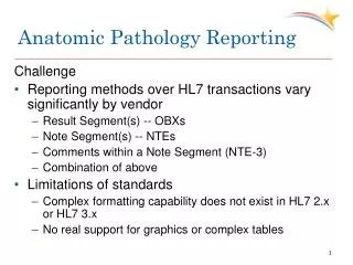
“Electronic” Requisition Forms • We do not get the information electronically • Scanning solutions must consider: • Workflow needs • Scanning speed • Appropriate/accurate labeling of files (scanned documents) to allow retrieval • Error handling • Availability of scanned documents for review by attendings • Long term storage of scanned documents John H. Sinard, MD, PhD
Document Archiving Solution • Create barcode labels and affix to documents • Scan documents • Use software to read barcode labels and rename documents • Resample/downsize the documents • Use existing image filing solution to file the documents in a separate file repository • Use existing image “Upload” software for manually addressing unreadable barcodes John H. Sinard, MD, PhD
Document Labeling Before Scanning • Want to label documents with barcodes • Requisition forms do not arrive pre-barcoded; significant outreach material • Would like to label with the accession number; not known in advance • Human readable as well as machine readable desired • Confidence in filing • Allows manual filing of unreadable barcodes • Solution: System-generated barcode “slide”-labels • Required creating a custom application object in CoPath • Can make on the fly; ability to make more • Can use existing label printers • Label size small enough not to obscure document • Barcode labels can also be used on other things (eg specimen containers) John H. Sinard, MD, PhD
Creating Document Labels in CoPath John H. Sinard, MD, PhD
Document Scanning • Document Scanning Requirements • Produce images of sufficient resolution to allow resolving small barcodes • Fast; automatic feed • Easy to use without training • Accommodate variability in paper weight, quality, and smoothness • Reliable; maintained (preferably by someone else) • Redundant and Multi-site • Solution: Upgraded our rented copiers to ones with scanning capabilities • Copier can create TIFF files (black and white) and scan to a networked drive • Small incremental cost • In some cases, needed to run new network lines John H. Sinard, MD, PhD
Labeling Reality John H. Sinard, MD, PhD
Requisition Image Filing • Automate the filing process: have software read the barcodes • Barcode Reading Software issues • Barcode size must accommodate 11 alphanumeric characters • Small barcode font requires high resolution label printer and scanner • Software needs to be “resistant” to variations in label placement and alignment • Solution: Custom Software • Scanned document (5100x6600 550KB TIFF) placed in a “Requisition Drop” folder • Folder action immediately detects file and runs a Perl script • Perl script uses Barcode Reading Software from Softek to read accession number • Also uses Image Processing software to down-sample the image into a 1000x1294 pixel, ~270KB JPEG image • New JPEG image file renamed to image file naming convention and placed into the Image Drop folder • Image filing engine files the document in the requisition repository John H. Sinard, MD, PhD
Ad hoc Images Image Filing Engine Image Repository Lab Info System Database Image Files Yale Pathology Clinical Imaging Intranet Access Manual Naming Gross Image Capture Photomicrographs Image Drop Folder Custom Image Uploading Program Electron Micrographs Images in Reports Step 1 Step 2 John H. Sinard, MD, PhD
Getting Images into the Image Drop Folder (Image File Naming Convention) To assure that images are uniquely identified, associated with the correct case, and readily located, a standardized naming convention is used for ALL CLINICAL IMAGES PPYYYY-AAAAAAcNN-dddd.fff File extension: image file format (should be .jpg) 1 or 2 Letter Specimen Prefix (eg, S, A, SB) 1 character lower case image type code 4 digit year [OPTIONAL] Dash plus up to 4 character (lower case) description abbreviation g: gross photograph m: photomicrograph c: cytology i: immunofluorescence e: electron micrograph r: imaging study f: FISH study d: document a: additional study u: unknown x: external report y: Yale Path report z: requisition form 6 digit accession number (with leading zeros) 2 digit sequential number (start with 01 for each type code) John H. Sinard, MD, PhD
Requisition Repository Image Filing Engine File Naming/ Resampling Engine Intranet Access Image Repository Barcoded Label S05-1234 Smith, John 01234567 Unreadable Barcodes Copier/Scanner Req Drop Folder Image Drop Folder CoPath Image Upload Program Image Files Database Yale Pathology Clinical Imaging Requisition Form Images in Reports Gross Images Photo-micrographs Electron Micrographs John H. Sinard, MD, PhD
Requisitions Available via Existing Web Interface John H. Sinard, MD, PhD
Outcome • We are currently accumulating: • ~2,500 images per month • ~5,000 requisition forms per month from Sept 2005 through Apr 2007 (Surg Path only) • >12,000 requisition forms per month since implementing in Cytology • ~ 4 document scans per day have to be manually filed because “the bar-code could not be read” • In most cases, the person scanning forgot to put the bar-code label on • No more sending of requisitions for microfilming • Unexpected benefit: requisitions readily available for responding to insurance denials • Have also created a final report repository John H. Sinard, MD, PhD
Thank you for your time and attention The Yale Pathology ITS Team Dr. Peter Gershkovich Aggie Daley János Löbb Katie Henderson Brian Paquin Sophia Gyory Garry Archer Wolfgang Freis Emma Walz-Vaitkus John H. Sinard, MD, PhD