Download
1 / 29
300 likes | 361 Views
Explore the structure and function of vertebrae, thorax, pelvis, and joints in the human body. Learn about the different types of joints and common inflammatory conditions associated with them. Gain insights into the clinical forms of arthritis.

E N D
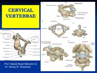
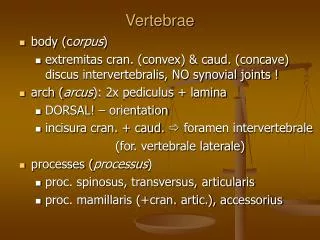
The Vertebral Column • Each vertebrae is given a name according to its location • There are 24 single vertebral bones separated by intervertebral discs of fibrocartilage • Seven cervical vertebrae are in the neck (C1-C7) • C1—Atlas • C2—Axis • Twelve thoracic vertebrae are in the chest region (T1-T12) • Five lumbar vertebrae are associated with the lower back (L1-L5)
The Vertebral Column Figure 5.14
A Typical Vertebrae, Superior View Figure 5.17
Regional Characteristics of Vertebrae Figure 5.18a
Sacrum and Coccyx • Sacrum • Formed by the fusion of five vertebrae • Coccyx • Formed from the fusion of three to five vertebrae • “Tailbone,” or remnant of a tail that other vertebrates have
Sacrum and Coccyx Figure 5.19
The Bony Thorax • Forms a cage to protect major organs • Consists of three parts • Sternum • Ribs • True ribs (pairs 1–7) • False ribs (pairs 8–12) • Floating ribs (pairs 11–12) • Thoracic vertebrae
The Bony Thorax Figure 5.20a
Bones of the Pelvic Girdle • Formed by two coxal (ossa coxae) bones • Composed of three pairs of fused bones • Ilium • Ischium • Pubis
Bones of the Pelvic Girdle • The total weight of the upper body rests on the pelvis • It protects several organs • Reproductive organs • Urinary bladder • Part of the large intestine
The Pelvis Figure 5.24a
The Pelvis: Right Coxal Bone Figure 5.24b
Gender Differences of the Pelvis • The female inlet is larger and more circular • The female pelvis as a whole is shallower, and the bones are lighter and thinner • The female ilia flare more laterally • The female sacrum is shorter and less curved • The female ischial spines are shorter and farther apart; thus the outlet is larger • The female pubic arch is more rounded because the angle of the pubic arch is greater
Joints • Articulations of bones • Functions of joints • Hold bones together • Allow for mobility • Ways joints are classified • Functionally • Structurally
Functional Classification of Joints • Synarthroses • Immovable joints • Amphiarthroses • Slightly moveable joints • Diarthroses • Freely moveable joints
Structural Classification of Joints • Fibrous joints • Generally immovable • Cartilaginous joints • Immovable or slightly moveable • Synovial joints • Freely moveable
Summary of Joint Classes [Insert Table 5.3 here] Table 5.3
Fibrous Joints • Bones united by fibrous tissue • Example: • Sutures • Syndesmoses • Allows more movement than sutures • Example: Distal end of tibia and fibula
Cartilaginous Joints • Bones connected by cartilage • Example: • Pubic symphysis • Intervertebral joints
Synovial Joints • Articulating bones are separated by a joint cavity • Synovial fluid is found in the joint cavity
Features of Synovial Joints • Articular cartilage (hyaline cartilage) covers the ends of bones • A fibrous articular capsule encloses joint surfaces • A joint cavity is filled with synovial fluid • Ligaments reinforce the joint
Structures Associated with the Synovial Joint • Bursae—flattened fibrous sacs • Lined with synovial membranes • Filled with synovial fluid • Not actually part of the joint • Tendon sheath • Elongated bursa that wraps around a tendon
The Synovial Joint Figure 5.29
Types of Synovial Joints Figure 5.30a–c
Types of Synovial Joints Figure 5.30d–f
Inflammatory Conditions Associated with Joints • Bursitis—inflammation of a bursa usually caused by a blow or friction • Tendonitis—inflammation of tendon sheaths • Arthritis—inflammatory or degenerative diseases of joints • Over 100 different types • The most widespread crippling disease in the United States
Clinical Forms of Arthritis • Osteoarthritis • Most common chronic arthritis • Probably related to normal aging processes • Rheumatoid arthritis • An autoimmune disease—the immune system attacks the joints • Symptoms begin with bilateral inflammation of certain joints • Often leads to deformities
Clinical Forms of Arthritis • Gouty arthritis • Inflammation of joints is caused by a deposition of uric acid crystals from the blood • Can usually be controlled with diet