Download
1 / 29
310 likes | 681 Views
A. Components of blood 1. Blood plasma 2. Formed elements B. Formation of blood cells C. Erythrocytes (red blood cells) 1. RBC anatomy 2. RBC physiology 3. RBC lifespan and number D. Leukocytes (white blood cells) 1. WBC anatomy and types

E N D
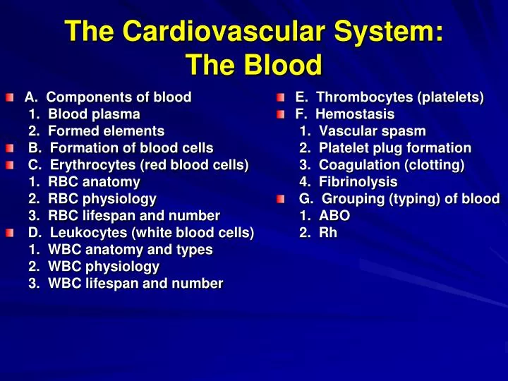
A. Components of blood 1. Blood plasma 2. Formed elements B. Formation of blood cells C. Erythrocytes (red blood cells) 1. RBC anatomy 2. RBC physiology 3. RBC lifespan and number D. Leukocytes (white blood cells) 1. WBC anatomy and types 2. WBC physiology 3. WBC lifespan and number E. Thrombocytes (platelets) F. Hemostasis 1. Vascular spasm 2. Platelet plug formation 3. Coagulation (clotting) 4. Fibrinolysis G. Grouping (typing) of blood 1. ABO 2. Rh The Cardiovascular System: The Blood
The more specialized a cell becomes to perform a specific function, the less capable it is of an independent existence. • As a result, it cannot: 1. protect itself 2. seek and procure food 3. move away from its own wastes
Internal Environment • Extracellular fluid (ECF) • a. interstitial fluid • b. plasma • c. lymph • 2. Intracellular fluid
Components of BloodFormed Elements (45%) • 1. erythrocytes (RBCs) • 2. leukocytes (WBCs) • 3. thrombocytes (platelets)
Components of Blood (Plasma ( 55%) Plasma Components A. water (91.5%) B. solutes (8.5%) (1) albumins (54-60%) (2) globulins (36-38%) (3) fibrinogen (4-7%)
Hematopoiesis • Yolk sac stage (3rd week – end of 2nd month) • Hepatic stage (beginning of 2nd month – just after birth) • Myeloid stage (beginning of 5th month – death)
Hematopoiesis (Hemopoiesis) hemocytoblast • 1. hemocytoblast • 2. stem cells • 3. growth factors a. erythropoietin b. thrombopoietin c. leukopoietins (?) myeloid stem cell lymphoid stem cell lymphocytes erythrocytes megakaryocytes neutrophils eosinophils basophils monocytes B cells T cells
Erythrocytes • 1. 7-10 um diameter • 2. biconcave disc • 3. large surface area • 4. very flexible • 5. not "true" cells • 6. sacs of hemoglobin • males = 4.6 – 6.2 million/mm3 hematocrit = 45-52% • females = 4.2 – 5.4 million/mm3 hematocrit = 37-48%
Hemoglobin • 1. 1 globin (protein) • 2. 4 Fe-containing heme groups • 3. 1 O2/heme • 4. 1 Hb = 4 O2 • 5. 1 globin = 1 CO2 • Hemoglobin value: Normal: Female 12-16 g/dL Male 13-18 g/dL
RBC Lifespan • 1. 120 days • 2. synthesis (2 million/sec) a. required substances b. erythropoietin regulation • 3. destroyed by liver and spleen (2 million/sec) • 4. recycled hemoglobin a. hemosiderin b. bilirubin c. amino acids
Erythrocyte Lifespan 120 days in circulation Synthesis (2 million/sec) decreased blood O2 kidneys release erythropoietin negative feedback stimulates required substances myeloid stem cells produce RBCs iron (Fe3+ ) globulin vitamin B12 erythropoietin increased blood O2 new RBCs liberated into bloodstream erythropoietin regulation Destroyed by liver and spleen (2 million/sec) RBCs destroyed Hb released broken down to Recycling of hemoglobin biliverdin hemosiderin globin + broken down to bilirubin Fe 3+ amino acids excreted recycled
Leukocytes (WBCs)= 5,000 – 10,000/mm3 • 1. granulocytes a. neutrophils (60 - 70%) phagocytize bacteria; 1st line of defense b. eosinophils (2 - 4%) phagocytize Ab-Ag complexes and allergens; anti-parasitic c. basophils (0.5 - 1%) secrete histamine and heparin • 2. agranulocytes d. lymphocytes (20 - 25%)- immunity e. monocytes (3 - 8%) become macrophages; phagocytosis; Ag-presentation
WBC Physiology • 1. chemotaxis • 2. chemotactic factors • 3. margination • 4. pavementing • 5. diapedesis • 6. emigration • 7. ameboid motion • 8. short lifespan 1. and 2. 7. 7. 5. and 6. 3. and 4.
Thrombocytes (Platelets) = 130,000 – 360,000/mm3 • 1. megakaryocyte • 2. cytoplasmic fragment • 3. not "true" cell • 4. biconvex disc • 5. 2 um diameter • 6. clotting factors • 7. lifespan 5 - 9 days
Hemostasis= Stoppage of Bleeding • Three basic mechanisms go into operation at the same time: • 1. vascular spasm • 2. platelet plug formation • 3. coagulation
Three Basic Mechanisms of Hemostasis 1. adhesion 2. release reaction 3. aggregation
Coagulation (Clotting) • 1. clot • 2. serum • 3. three basic processes a. prothrombinase b. prothrombin --> thrombin c. fibrinogen --> fibrin • 4. clot retraction (syneresis) Prothrombinase Fibrinogen
Fibrinolysis = dissolution of a clot • Plasminogen activating factor (kallikrein) (converts) plasminogen plasmin clot dissolved clot
Grouping (Typing of Blood) • 1. glycolipid agglutinogens • 2. two major blood group systems a. ABO b. Rh
Blood Types • Membrane Bound Agglutinogens (Antigens) ABO Blood System and Rh System • Plasma antibodies (agglutinins) (Anti A and Anti B- no anti Rh) Cause agglutination and hemolysis Type A Type B Type AB Type O RBCs Agglutinogen A Agglutinogen B Agglutinogens A and B No Agglutinogens Plasma Neither Agglutinin A or B Both Agglutinin A and B Agglutinin B Agglutinin A
Who can give what to whom? Recipient is type A. Can she receive: • type A plasma • type A RBCs • type B plasma • type B RBCs • type AB plasma • type AB RBCs • type O plasma • type O RBCs
Who can give what to whom? Recipient is type B. Can she receive: • type A plasma • type A RBCs • type B plasma • type B RBCs • type AB plasma • type AB RBCs • type O plasma • type O RBCs
Who can give what to whom? Recipient is type AB. Can she receive: • type A plasma • type A RBCs • type B plasma • type B RBCs • type AB plasma • type AB RBCs • type O plasma • type O RBCs
Who can give what to whom? Recipient is type O. Can she receive: • type A plasma • type A RBCs • type B plasma • type B RBCs • type AB plasma • type AB RBCs • type O plasma • type O RBCs
Rh System of Grouping • 1. agglutinogen Rh • 2. no agglutinin _________________ • hemolytic disease of the newborn • (erythroblastosis fetalis)