Download
1 / 60
600 likes | 612 Views
Explore the spectrum of benign and malignant stomach diseases, from acute gastritis to peptic ulcers, and learn about their diagnosis and treatment with in-depth insights from Prof. Dr. Öge Taşcılar. Delve into topics such as Helicobacter pylori infection, peptic ulcer disease complications, neoplasms of the stomach, and more.

E N D
Benignandmaligndiseases of Stomach Prof. Dr. Öge TAŞCILAR
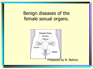
MİDE • Mukoza • Submukoza • MuskularisPropria • Seroza • Mukoza İntraepitelyal Mukoza Epitel Bazal Membran LaminaPropria Muskularis Mukoza
MİDE • Fonksiyon: • Alınan gıdaların sindirimi ve emilimi • Reseptifrelaksasyon ve gastrik adaptasyon • İntragastrik basınç düşer. • 100cc-------------1500cc
MİDE • Mallory-Weiss Sendromu • Kusma ile ÖG bileşke mukoza submukoza yırtık ve kanama • Endoskopi • Alkol,diyabet,gebelik, üremi, • Tam kat olursa Booerhaave sendromu
MİDE • Bezoarlar • Midede oluşan yabancı cisimler. • Trikobezoar-fitobezoar • Mide operasyonu sonrası • Antrumun öğütücü işlevinin kaybolması • HCL azalmasına bağlı CandidaAlbicansbezoar • Tanı: Radyoloji-endoskopi • Tedavi:Endoskopik-Cerrahi
MİDE • Menetrier Hastalığı • Hipertrofikmukozalgastropati • Fundus ve korpusta dev rugalar • Foveolarhiperplazi • Hipoklorhidri ve Hipoalbüminemi • 50> erkekler • Epigastrik ağrı, kilo kaybı, (özellikle protein) , kanama, diare, ödem • Medikal tedavi PPI • Destek tedavisi • Çok ciddi olgularda rezeksiyon
Acute gastritis • Drugs (non-steroidal anti-inflammatory drugs NSAID), alcohol cause acute erosion (loss of mucosa superficial to muscularis mucosae). Can result in severe haemorrhage
Chronic gastritis ABC • A – autoimmune(associated with vitamin B12 malabsorption (pernicious anaemia) • B – bacterial (helicobacter) • C – chemical(bile reflux, drugs)
Autoimmune chronic gastritis • Autoantibodies to gastric parietal cells • Hypochlorhydria/achlorhydria • Loss of gastric intrinsic factor leads to malabsorption of vitamin B12 with macrocytic,megaloblastic anaemia
Helicobacter pylori • Adapted to live in association with surface epithelium beneath mucus barrier • Causes cell damage and inflammatory cell infiltration • In most countries the majority of adults are infected
Peptic ulcer disease • A surface breach of mucosal lining of GI tract occurring as a result of acid and pepsin attack • Sites: • Duodenum (DU) • Stomach (GU) • Oesophagus • Gastro-enterostomy stoma • Related to ectopic gastric mucosa (e.g. in Meckel’s diverticulum)
Pathogenesis • In normal acid/pepsin attack is balanced by mucosal defences • Increased attack by hyperacidity • Weakened mucosal defence – the major factor (H. pylori related)
MİDE • Duodenal Ülser: • Duodenal HCO3 sekresyonu azalmış • Gece asit sekresyonu artmış • Duodenal asit yükü artmış • Bazal ve postprandial gastrin artmış • Tamamına yakın HP gastrit saptanmıştır.
Morphology of peptic ulcers • Clean, non-elevated edge • Granulation tissue base (floor) • Underlying fibrosis
MİDE • Klinik: • Yanıcı, kemirici, açlık ağrısı. • Epigastrium • Antiasit ve gıda ile hafifler. • Mevsimsel bir ağrı. • İlkbahar, sonbahar, stress dönemleri • Penetre olursa ağrı özellikleri değişir.
MİDE • Anamnez, • Radyoloji • Endoskopi, biyopsi • Tedavi: • Medikal tedavi Antiasit Sükralfat H2 blokör PPI Prostoglandin analogları
MİDE • DÜ Cerrahi Tedavi: • BTV-PP • BTV-Distal gastrektomi+GJ • PGV
MİDE • Mide Ülseri
Complications of peptic ulcer • Perforation leading to peritonitis • Haemorrhage by erosion of vessel in base • Penetration of surrounding organ (liver/pancreas) • Obstruction (by scarring) – pyloric stenosis • (Cancer – rare event in true peptic ulcer)
NEOPLASMS OF STOMACH BENIGN__ 10% MALIGNANT__90% BENIGN Polyps Lipomas Leiomyomas
NEOPLASMS OF STOMACH MALIGNANT Adenocarcinoma95% Lymphoma4% Others1% (sq.cellca, angiosarcoma,carcinosarcoma, Gist)
Less common gastric neoplasms • Gastrointestinal stromal tumour (GIST) • Lymphoma • Neuroendocrine (carcinoid) tumours
GIST • Risk categories were assigned according to current recommended NIH criteria. • Tumors <2 cm and<5 mitosis per 50 high-power fields (HPF) were classified as very low risk. • Tumors ranging from 2 to 5 cm and having <5 mitoses/50 HPF were classified as low risk.
Tumors <5 cm but having 6 to 10 mitoses/50 HPF were intermediate risk, as were tumors from 5 to 10 cm with <5 mitoses/50 HPF. • Tumors >5 cm with >5 mitoses/50 HPF was defined as high risk, as was any tumor >10 cm or any tumor with >10 mitoses/50 HPF.
GASTRIC STROMAL TUMOURS PRESENTATION; Mass abdomen Upper GI bleeding Obstruction PATHOLOGY; Difficult to ascertain benign or malignant nature Size & Histology is the criteria TREATMENT; Surgical resection Lymph node resection not necessary.
MİDE LENFOMA • Non-HodgkinLenfoma(NHL) klasik olarak lenf nodlarından gelişir. • Ama NHL %30 olguda ekstranodal(Solid organ kaynaklı) olarak gelişebilir. • GI sistem tüm NHL %20
MİDE LENFOMA • GI lenfoma; oral kaviteden rektuma • En sık; Mide • Sonra ince barsak • Kolon • Pankreas
MİDE LENFOMA • NHL, ekstranodal lenfoma ve GI lenfomanın en sık görülen tipi diffüz B hücre lenfoması. • MALT lenfoma • Burkitt lenfoma • T- hücre lenfoma
Gastric lymphoma • Malignant neoplasm of mucosa associated lymphoid tissue (MALT) • A (usually) low grade B-cell (marginal cell) lymphoma
MİDE LENFOMA • Antrum ve distal mide • Proksimal yerleşebilir. • Karın ağrısı, erken doyma • Bulantı, kusma, halsizlik • Abdominal dolgunluk • Kronik kan kaybı, anemi melena
Gastric lymphoma (maltoma) • Neoplastic cells infiltrate the epithelium (lymphoepithelial lesions) • Strongly associated with chronic H. pylori and can be cured by eliminating infection.
MİDE LENFOMA • Tedavi • HP tedavi edilmeli. • Bir zamanlar cerrahi • Şimdi Konservatif, bazı olgularda cerrahi • Low grade lenfoma(MALT) • HP eradikasyonu, KRT, • High Grade: • Antihelikobakter tedaviye cevap vermeyen erken evre PGL, ileri evre lenfoma, diffüz büyük hücreli lenfoma ise cerrahi tedavi • KT-RT • Residual hastalık: KT-cerrahi
Neuroendocrine tumours • Carcinoids are tumours of resident neuroendocrine cells in gastric glands • Usually seen in context of chronic atrophic gastritis (driven by gastrin) • Clinical behaviour variable
ETIOLOGY 1.HELICOBACTER PYLORI CA of body & distal stomach Gastritis Gastric atrophy Intestinal metaplasia 2.PERNICIOUS ANEMIA 3.GASTRIC POLYPS 4.Pt. with surgery of peptic ulcer disease Billroth II gastrectomy Gastroenterostomy Pyloroplasty 4 times increased risk
5.Cigarette smoking &dust ingestion 6.Diet Consumption of potatoes,pickledvegetables,dried/salted fish & less milk Alcohol ingestion Excessive salt intake Deficiencies of anti oxidants Exposure to N- Nitrosocompounds 7.Familial predisposition Relatives of CA stomach pt. are 4 times more at risk Genetically H-ras, C-erb B2 & APC gene mutations have some role in pathogenesis of CA stomach Blood group A 8.Gastric ulcer 3-5% of cases?? 9. İntestinalmetaplazi Tip1,2 ve 3 En tehlikeli olanı Tip 3
PATHOLOGY MACROSCOPIC CLASSIFICATION Schirrous (lintis plastica) Ulcerative Polypoid Superficial spreading HISTOLOGICALLY (W.H.O) Papillary Tubular Mucin secreting Signet ring cell
Clinical Features GASTRIC CANCER Early feeling of fullness after meal Bloating , distention Vomiting Pallor – iron deficiency anemia due to tumour bleed Dysphygia –epigastric fullness or vomiting due to obstrution of gastric outlet Epigastric mass – ¼ of cases Non metastatic effects ; thrombophlebitis Deep venous thrombosis ( by affecting thrombotic & haemostatic mechanism)
Ascites Jaundice Trosier sign(virchows node) Sister mary joseph nodule krukenbergtumour Blummer Shelf
INVESTIGATIONS BLOOD COMPLETE EXAM. ------ Anemia STOOL EXAM. --- for occult blood in ½ of pts. CARCINOEMBRYONIC (CEA) LEVEL--- elevated in 65% of cases GASTRIC JUICE ANALYSIS--- 20% are achlorhydric after maximal stimulation DOUBLE CONTRAST BARIUM MEAL--- mucosal irregularities and to assess the size , shape, margins of lesions GASTROSCOPY & BIOPSY---minimum of 6 biopsies for accuracy --- brush cytology C.T. SCAN ENDOSCOPIC USG LAPAROSCOPY