Download
1 / 50
530 likes | 830 Views
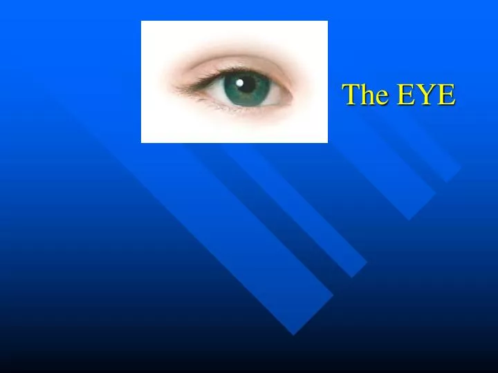
The EYE. Vision. Dominant sense in humans Performed by eyes , in orbits of skull Surrounded by accessory structures Positioned by six extrinsic muscles. Accessory Eye Structures. Eyebrows Eyelids (Palpebrae) Eyelashes Conjunctiva Lacrimal glands/ducts Extrinsic eye muscles.

E N D
Vision • Dominant sense in humans • Performed by eyes, in orbits of skull • Surrounded by accessory structures • Positioned by six extrinsic muscles
Accessory Eye Structures • Eyebrows • Eyelids (Palpebrae) • Eyelashes • Conjunctiva • Lacrimal glands/ducts • Extrinsic eye muscles
Functions of Accessory Structures: • Eyebrows – • physical protection • Eyelashes – • blinking reflex initiation • Eyelids – • lubrication • contain sebaceous glands
CONJUNCTIVA • transparent mucous membrane • lines inner surface of eyelids • Folds over / covers eyeball surface • MAJOR LUBRICATION FUNCTION!
The Lacrimal Apparatus (gland + ducts)!! • Location: superior / lateral to eyeball • Continuous lacrimal secretion ! (aka tears) • Blinking: spreads lacrimal secretions over eyeball surface • Lacrimal canals (medial to eye) drain secretion to lacrimal sac… • Lacrimal sac to nasolacrimal duct to nasal cavity!
Extrinsic Eye Muscles • Move eyeball • Six (6) per eye! • LATERAL RECTUS • MEDIAL RECTUS • SUPERIOR RECTUS • INFERIOR RECTUS • INFERIOR OBLIQUE • SUPERIOR OBLIQUE
Reminder: What Cranial Nerves control these muscles? III, IV, VI !!
The Eyeball • Sphere of three layers (TUNICS) FIBROUS TUNIC (Outermost) VASCULAR TUNIC (Middle layer) SENSORY TUNIC (Innermost)
Fibrous Tunic • CT • Two major Regions: 1. SCLERA : • The “white” of the eye • Durable, provides shape to the eyeball ant. • Point of attachment for extrinsic eye muscles • Continuous with Dura Mater • Surrounds the Optic Nerve (II) post.
2. CORNEA • Most anterior portion of fibrous tunic • Continuous with sclera • Transparent • “window” of the eye
Vascular Tunic • blood vessels • Three Major Regions: • Choroid • Ciliary Body • Iris
CHOROID • Structure: • Located posteriorly • Dark brown pigmentation (melanin) • Functions: • Provides nutrition to all tunics! • Melanin absorbs light (prevents reflection)
CILIARY BODY • Structure: • Thick ring of : • ciliary muscles and suspensory ligaments • Ciliary processes (^ capillaries) • Functions: • Surround/control shape of lens • Secrete fluid found in anterior chamber of eyeball
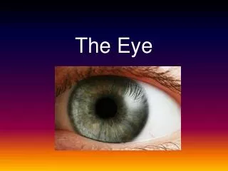
IRIS • Structure: • Most anterior portion of Vascular Tunic • Located between cornea and lens • Flattened appearance • Contain smooth muscle fibers • Surrounds the PUPIL (central opening that allows light to enter the eye!) • Contains MELANIN • ^ melanin = brown eyes • melanin = green / blue /non brown eyes
Functions: • Controls diameter of the PUPIL! • Reflexive to: • Amount of Light present! • Distance of object being seen! • Emotional responses!
Pupil Constriction (PC) or Pupil Dilation (PD)??? • Reading a book 6 inches from eyes? • PC • Looking at a traffic sign while driving? • PD • Solving a mathematical word problem? • PD • Seeing something attractive approaching? • PD • Seeing something repulsive or boring approaching? • PC • Having a flashlight shown directly at face? • PC • Entering a dark room? • PD
Sensory Tunic • Aka… RETINA • Structure: • 2 Layers: • Outer Pigmented Layer • Absorbs light/ prevents scattering & reflection • Stores Vitamin A!! • Inner Nervous Layer • Contains Photoreceptor Cells! Neurons + • Contains Optic Nerve neuroglia!
The Eye and Vision choroid sclera retina
Retina cont’d. • Function of the Retina • Complex explanation: • Processes light stimuli and sends impulses to brain via the optic nerve! • Simple explanation: • ALLOWS YOU TO SEE!
Retina continued… • So What about these Photoreceptor Cells? • 2 TYPES: • RODS receptors for dim light and peripheral vision! • CONES receptors for bright light and color!
Absorb all wavelengths of visible light ~200 rods relay impulse to single neuron of OPTIC nerve Widely dispersed in retina; allows for greater field of vision Absorb Red, Green, Blue wavelengths 1 cone to 1 OPTIC nerve neuron impulse ratio! Allows for better visual acuity! Rods vs. Cones
Vision in a Nutshell… • Rods and Cones receive light stimuli and generate impulse!! • Impulse passed to Bipolar Neurons! • Impulse passed to Ganglion Neurons! • Action Potential generated along OPTIC NERVE!!
Structures Associated with the Retina • OPTIC DISC • Area where optic nerve exits the eye! • Very weak… no support from sclera! • Lacks photoreceptors! (uh-oh…) • Aka Blind Spot
Central Artery • Central Vein • Transport blood to / from retina • Enter/leave eye through center of Optic Nerve • Form highly branched network of vessels!
The LENS Structural Characteristics: Biconvex! Transparent! Avascular! (Why??) Flexible! Held in place and changes position due to… Suspensory ligaments !!
Function of Lens: • Adjusts shape to focus light on the retina!! • Divides Eye into Anterior and Posterior Chambers!
Lens Problems … • As body ages lens becomes… • Thicker • More convex • Less flexible THE RESULT OF LENS PROBLEMS…??
Internal Chambers of the Eye • Lens is the DIVIDING LANDMARK!! • Anterior Chamber: • Filled with Aqueous Humor • Watery, thin, clear fluid • Constantly produced and drained • Posterior Chamber: • Filled with Vitreous Humor • Thick, clear gel • Formed embryonically… lasts for lifetime
vitreous humor lens aqueous humor